

Photodynamic therapy and cell death pathways
- PMID: 20552338
- PMCID: PMC4455965
- DOI: 10.1007/978-1-60761-697-9_3
Photodynamic therapy and cell death pathways
Abstract
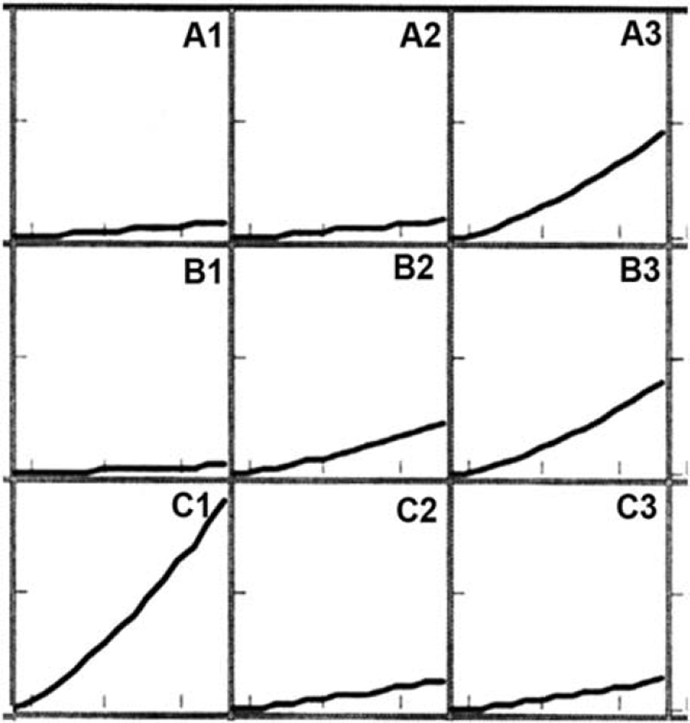
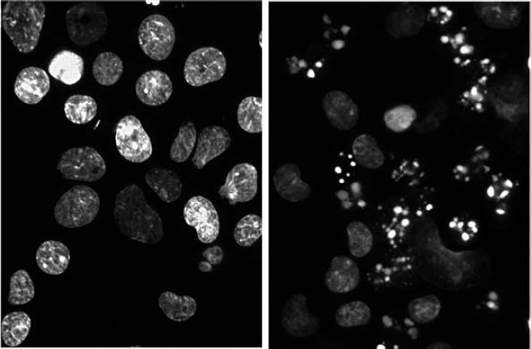
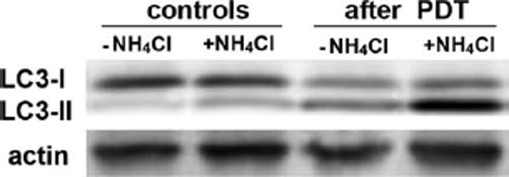
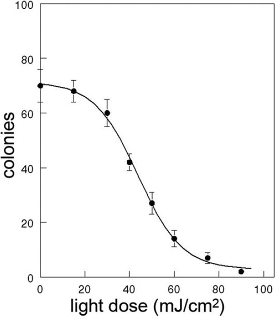
Photodynamic therapy (PDT) is the term used to describe the irradiation of photosensitized cells or tissue with phototoxic consequences. This process can result in the rapid initiation of not only apoptosis, an irreversible death pathway, but also autophagy. The procedures described here are designed to characterize the correlation between the PDT dose vs. survival of cells in vitro, the apoptotic effects of photodamage, and the extent of an autophagic response. These are assessed by clonogenic assays, observation of condensed chromatin characteristic of apoptosis, activation of "executioner" caspases, and the autophagic flux as indicated by comparing accumulation of the LC3-II protein under conditions where processing of autophagosomes is retarded vs. is not retarded.
Figures
References
-
- Kessel D, Castelli M. Evidence that Bcl-2 is the target of three photosensitizers that induce a rapid apoptotic response. Photochem Photobiol. 2001;74:318–322. - PubMed
-
- Xue LY, Chiu SM, Oleinick NL. Photochemical destruction of the Bcl-2 oncoprotein during photodynamic therapy with the phthalocyanine photosensitizer Pc 4. Oncogene. 2001;20:3420–3427. - PubMed
-
- Xue LY, Chiu SM, Fiebig A, Andrews DW, Oleinick NL. Photodamage to multiple Bcl-xL isoforms by photodynamic therapy with the phthalocyanine photosensitizer Pc 4. Oncogene. 2003;22:9197–9204. - PubMed
Publication types
MeSH terms
Substances
Grants and funding
LinkOut - more resources
Full Text Sources
