. 2010 Nov;131(3):426-37.
doi: 10.1111/j.1365-2567.2010.03316.x.

Evidence that CD8 T-cell homeostasis and function remain intact during murine pregnancy

Affiliations

Evidence that CD8 T-cell homeostasis and function remain intact during murine pregnancy

Michelle T Norton et al. Immunology. 2010 Nov.

Abstract

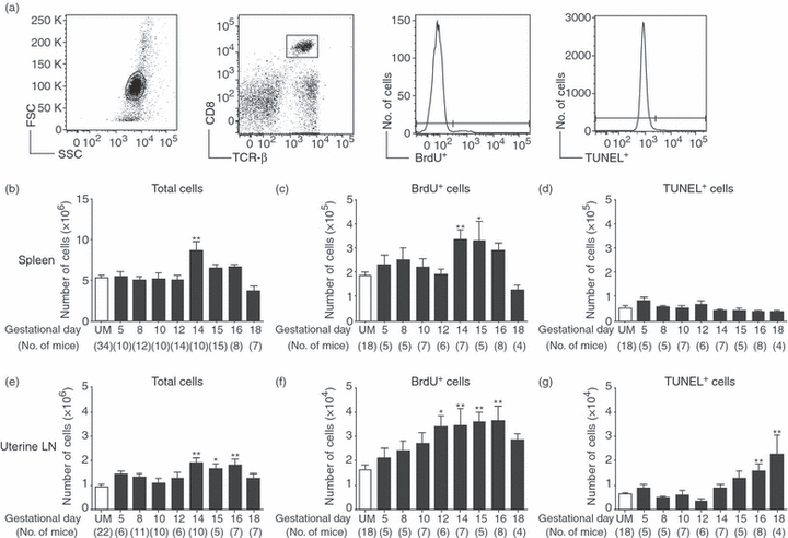
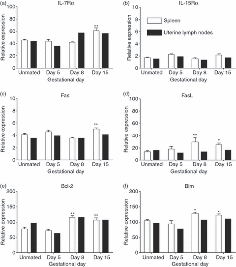
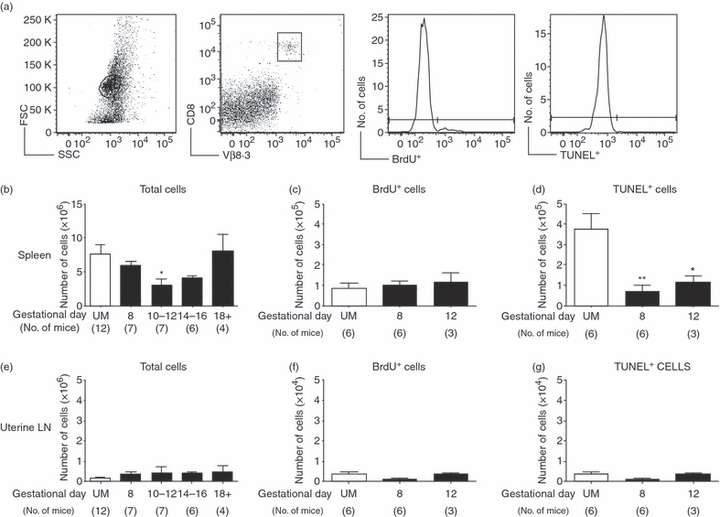
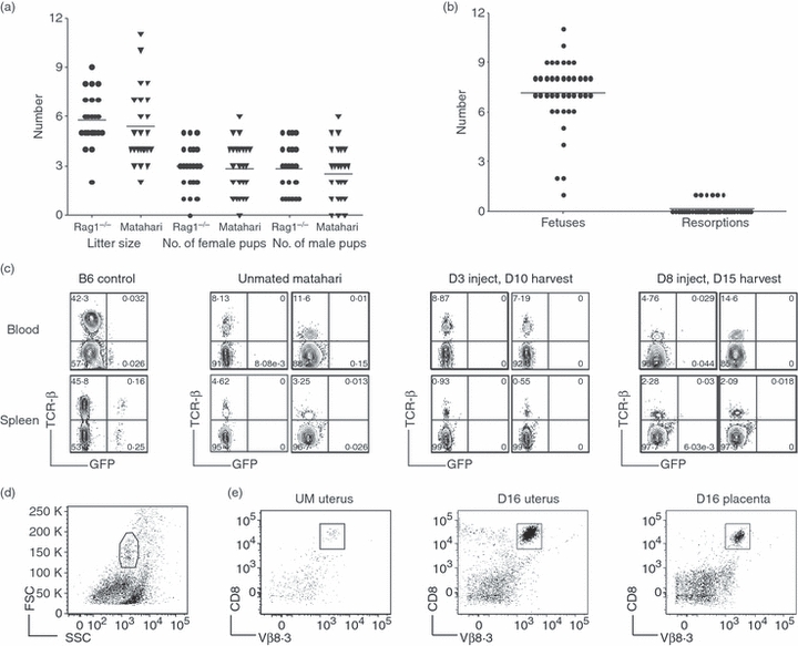
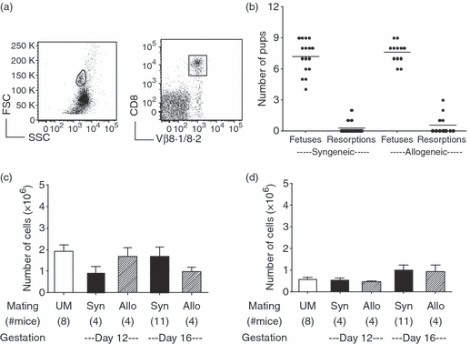
Evolving models of immune tolerance have challenged the view that the response of the maternal immune system to environmental or fetal antigens must be suppressed or deviated. CD8 T cells play a central role in the immune response to viruses and intracellular pathogens so the maintenance of both the number and function of these cells is critical to protect both the mother and fetus. We show that the numbers of maternal CD8 T cells in both the spleen and the uterine draining lymph nodes are transiently increased at mid-gestation and this correlates with enhanced CD8 T-cell proliferation and an increased relative expression of both pro-survival and pro-apoptotic molecules. In transgenic mice bearing T-cell antigen receptors specific for the male HY or allo-antigens, the transgenic CD8 T cells retain the ability to proliferate and function during pregnancy. Moreover, anti-HY T-cell receptor transgenic mice have normal numbers of male pups despite the presence of CD8 T cells at the maternal-fetal interface. These data suggest that pregnancy is a dynamic state in which CD8 T-cell turnover is increased while the function and ending size of the CD8 T-cell compartment are maintained.


Figures

Figure 1
Figure 1
Increased proliferation of CD8 T cells during normal pregnancy. Pregnant and unmated (UM) C57BL/6 mice were injected with bromodeoxyuridine (BrdU) for the 24 hr before death, and parallel samples from each mouse were analysed for proliferation by BrdU incorporation, and apoptosis by the terminal deoxynucleotidyl-transferase (TdT)-mediated dUTP nick-end labelling (TUNEL) assay. (a) Representative analysis of flow cytometric data from a day 12 pregnant mouse. Left to right: forward versus side scatter plot of whole spleen, gating of CD8+ TCR-β+ cells, histogram analysis for the proportion of BrdU+ cells, and histogram analysis for the proportion of TUNEL+ cells. (b) Numbers of CD8 T cells in the spleen were calculated by multiplying the total number of cells counted by the proportion of CD8+ TCR-β+ cells obtained by flow cytometry. (c) Number of splenic CD8+ T cells that were BrdU+ on each gestational day. (d) Number of splenic CD8+ T cells positive for TUNEL staining throughout pregnancy. (e) Numbers of CD8 T cells in the uterine draining lymph nodes were calculated by multiplying the total number of cells counted by the proportion of CD8+ TCR-β+ cells obtained by flow cytometry. (f) Number of uterine draining lymph node CD8+ T cells that were BrdU+ on each gestational day. (g) Number of uterine lymph node CD8+ T cells positive for TUNEL staining throughout pregnancy. In each graph symbols and error bars depict mean and standard error of the mean (SEM). Numbers in parenthesis represent the total number of mice analysed at each time-point. Statistical significance was determined by one-way analysis of variance with Dunnett's post-test using UM controls for comparison *P < 0·05, **P < 0·01.
Figure 2
Figure 2
Increased expression of cytokine receptor and pro-apoptotic genes during pregnancy. CD8 T cells were sorted using fluoresence-activated cell sorting from single spleens or pooled uterine draining lymph nodes (two or three mice) from unmated (UM) controls and day 5, 8 and 15 pregnant mice. RNA was prepared by Triazol extraction and analysed by quantitative reverse transcription polymerase chain reaction. Relative expression was determined by standard curve analysis and normalized by dividing by the geometric mean of two housekeeping genes. Shown is the relative expression of (a) interleukin-7 receptor α-chain (IL-7Rα), (b) IL-15Rα, (c) Fas, (d) Fas ligand (FasL), (e) Bcl-2, and (f) Bim in CD8 T cells from the spleen and uterine draining lymph nodes during pregnancy. Open bars represent spleen cells and filled bars are pooled uterine lymph node cells. Statistical significance of spleen samples was determined by one-way analysis of variance with Dunnett's post-test using UM controls for comparison *P < 0·05, **P < 0·01.
Figure 3
Figure 3
CD8 T-cell numbers in anti-HY T-cell receptor transgenic Matahari mice are maintained during pregnancy. Pregnant and unmated (UM) Matahari mice were injected with bromodeoxyuridine (BrdU) for the 24 hr before euthanasia, and parallel samples from each mouse were analysed for proliferation by BrdU incorporation, and apoptosis by the terminal deoxynucleotidyl-transferase (TdT)-mediated dUTP nick-end labelling (TUNEL) assay. In this system, transgenic CD8 T cells are identified by the Vβ8.3 T-cell receptor (TCR), and all analysis was performed on CD8+ Vβ8.·3+ cells. Cells positive for Vβ8.3 but negative for CD8 were rare and were excluded from our analysis. (a) Representative flow cytometry showing forward- versus side-scatter plot of whole spleen, gating of CD8+ Vβ8.3+ T cells, histogram analysis for the proportion of BrdU+ cells, and histogram analysis for the proportion of TUNEL+ cells. (b) Numbers of CD8 T cells in the spleen of Matahari mice were calculated by multiplying the total number of cells counted by the proportion of CD8+ Vβ8.3+ T cells obtained by flow cytometry. (c) Number of splenic CD8+ Vβ8.3+ T cells that were BrdU+ in UM and pregnant mice on days 8 and 12 of gestation. (d) Number of splenic CD8+ Vβ8.3+ T cells positive for TUNEL staining in UM mice and on days 8 and 12 of pregnancy. (e) Numbers of CD8+ Vβ8.3+ T cells in the uterine draining lymph nodes of Matahari mice were calculated by multiplying the total number of cells counted by the proportion of CD8+ Vβ8.3+ T cells obtained by flow cytometry. (f) Number of uterine lymph node CD8+ Vβ8.3+ T cells that were BrdU+ in UM and pregnant mice on days 8 and 12 of gestation. (g) Number of uterine lymph node CD8+ Vβ8.3+ T cells positive for TUNEL staining in UM mice and on days 8 and 12 of pregnancy. In each graph symbols and error bars depict mean and standard error of the mean (SEM). Numbers in parenthesis represent the total number of mice analysed from each time-point. Statistical significance was determined by one-way analysis of variance with Dunnett's post-test using UM controls for comparison *P < 0·05, **P < 0·01.
Figure 4
Figure 4
Anti-HY Matahari T-cell receptor (TCR) transgenic CD8 T cells are functional during pregnancy with no adverse effects on fetal survival. (a) Litter size and the number of male and female pups on weaning from breeding pairs of anti-HY Matahari TCR transgenic and non-transgenic Rag1−/− mice. (b) The number of fetuses and resorptions from 40 Matahari pregnancies at various gestational days. (c) CD8 T-cell activity was tested by injecting day 3 or 8 pregnant Matahari, unmated Matahari, and B6 control mice with male GFP+ spleen cells. Seven days later the presence of GFP+ cells was determined in the blood (top) and spleen (bottom) by flow cytometry. Non-transgenic B6 mice were used as controls (left most panels). Profiles for two mice from each time-point analysed are shown. Numbers refer to percentage of cells in the lymphocyte gate. (d) Representative flow cytometric gating from a Matahari day 16 uterus. (e) Isolation of CD8+ T cells from the uterus and placenta of Matahari mice. Fetal–placental units were removed and the uteri of four pregnant mice were pooled. Pooled uteri from four unmated mice were run as controls. For each pregnant mouse, two placentas from each uterine horn were pooled together (four in total). CD8 T cells were isolated from enzymatically digested uterus and placenta samples by magnetic antibody cell sorting positive selection and visualized by flow cytometry. CD8+ Vβ8.3+ T cells were purified from unmated uterus (left), day 16 uterus (middle), and day 16 placenta (right).
Figure 5
Figure 5
Allogeneic pregnancy does not alter the number of CD8 T cells in anti-Ld 2C transgenic mice. T-cell receptor (TCR) transgenic 2C Rag1−/− female mice were mated to either B6 Rag1−/− males for syngeneic pregnancies or to Ld expressing BALB/c Rag1−/− males for allogeneic pregnancies. Mice were analysed on gestational days 12 and 16 and compared with unmated controls. (a) Representative gating is shown from a day 12 pregnant mouse using CD8 and the clonotypic TCR Vβ8.1/8.2. (b) The number of fetuses and resorptions were counted in syngeneic and allogeneic 2C Rag1−/− pregnancies. (c) Numbers of CD8+ Vβ8.1/8.2+ T cells in the spleen. (d) Numbers of CD8+ Vβ8.1/8.2+ T cells in the uterine draining lymph nodes. Statistical significance was tested by one-way analysis of variance with Newman–Keuls post-test.

Similar articles

Cited by

References

    1. Baines M, Pross H, Millar K. Effects of pregnancy on the maternal lymphoid system in mice. Obstet Gynecol. 1977;50:457–61. - PubMed
    1. Hamilton M, Hellström I. Altered immune responses in pregnant mice. Transplantation. 1977;23:423–30. - PubMed
    1. Parmiani G, Invernizzi G. Depression of cellular immune response during syngeneic pregnancy as measured by the graft-versus-host reaction. Transplantation. 1975;19:363–8. - PubMed
    1. Jiang S, Vacchio M. Multiple mechanisms of peripheral T cell tolerance to the fetal “allograft”. J Immunol. 1998;160:3086–90. - PubMed
    1. Tafuri A, Alferink J, Möller P, Hämmerling G, Arnold B. T cell awareness of paternal alloantigens during pregnancy. Science. 1995;270:630–3. - PubMed

Publication types

MeSH terms

Substances

LinkOut - more resources