. 2010 Jun 16:7:17.
doi: 10.1186/1742-9994-7-17.

Confocal analysis of nervous system architecture in direct-developing juveniles of Neanthes arenaceodentata (Annelida, Nereididae)

Affiliations

Confocal analysis of nervous system architecture in direct-developing juveniles of Neanthes arenaceodentata (Annelida, Nereididae)

Christopher J Winchell et al. Front Zool. .

Abstract

Background: Members of Family Nereididae have complex neural morphology exemplary of errant polychaetes and are leading research models in the investigation of annelid nervous systems. However, few studies focus on the development of their nervous system morphology. Such data are particularly relevant today, as nereidids are the subjects of a growing body of "evo-devo" work concerning bilaterian nervous systems, and detailed knowledge of their developing neuroanatomy facilitates the interpretation of gene expression analyses. In addition, new data are needed to resolve discrepancies between classic studies of nereidid neuroanatomy. We present a neuroanatomical overview based on acetylated alpha-tubulin labeling and confocal microscopy for post-embryonic stages of Neanthes arenaceodentata, a direct-developing nereidid.

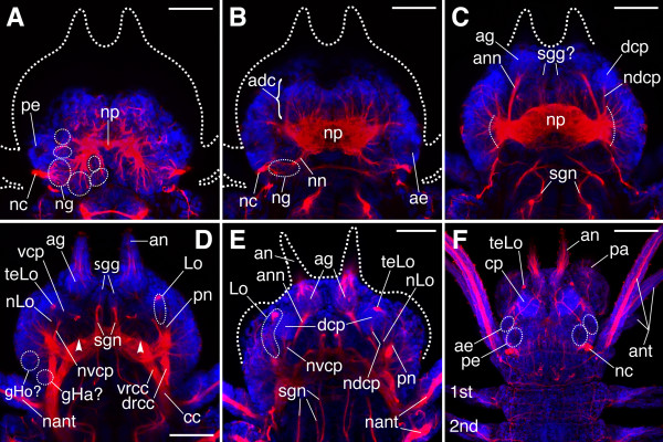
Results: At hatching (2-3 chaetigers), the nervous system has developed much of the complexity of the adult (large brain, circumesophageal connectives, nerve cords, segmental nerves), and the stomatogastric nervous system is partially formed. By the 5-chaetiger stage, the cephalic appendages and anal cirri are well innervated and have clear connections to the central nervous system. Within one week of hatching (9-chaetigers), cephalic sensory structures (e.g., nuchal organs, Langdon's organs) and brain substructures (e.g., corpora pedunculata, stomatogastric ganglia) are clearly differentiated. Additionally, the segmental-nerve architecture (including interconnections) matches descriptions of other, adult nereidids, and the pharynx has developed longitudinal nerves, nerve rings, and ganglia. All central roots of the stomatogastric nervous system are distinguishable in 12-chaetiger juveniles. Evidence was also found for two previously undescribed peripheral nerve interconnections and aspects of parapodial muscle innervation.

Conclusions: N. arenaceodentata has apparently lost all essential trochophore characteristics typical of nereidids. Relative to the polychaete Capitella, brain separation from a distinct epidermis occurs later in N. arenaceodentata, indicating different mechanisms of prostomial development. Our observations of parapodial innervation and the absence of lateral nerves in N. arenaceodentata are similar to a 19th century study of Alitta virens (formerly Nereis/Neanthes virens) but contrast with a more recent study that describes a single parapodial nerve pattern and lateral nerve presence in A. virens and two other genera. The latter study apparently does not account for among-nereidid variation in these major neural features.


Figures

Figure 1
Figure 1
SEM of juvenile developmental stages and basic external morphology of N. arenaceodentata. These worms were reared at ≈21°C, and all ages are approximate. Scale bars: A-I = 100 μm; H = 4 mm. Specimens processed for SEM experienced approximately 30% shrinkage. A is a lateral view, anterior to the left and ventral down; B, C, D, and G are ventral views, anterior to the left; E is a ventrolateral view, anterior to the upper left; F, I, and J are dorsal views with anterior up in I, and to the upper left in F and J. A. Hatchling, 10 days post-fertilization (dpf). B. Mid 3-chaetiger stage, 11 dpf. C. Late 3-chaetiger stage, 12 dpf. D. 6 chaetigers, 15 dpf. E. 9 chaetigers, 17 dpf. F. 13 chaetigers, 22 dpf. G. 20 chaetigers, 30 dpf. H. Transverse view (anterior side) of an adult parapodium taken from a mid-body segment. I and J are light micrographs. I. 4 chaetigers, 13 dpf. The developing pharynx underlies the asterisk. J. Adult, 70 dpf. 1st, 2nd, 3rd nascent parapodia, an antenna, ana anal cirrus, ant anterior cirrus, dc dorsal cirrus, dnl dorsal notopodial ligule, mo mouth, NE neuropodium, nec neurochaetae, nel neuropodial ligule, NO notopodium, noc notochaetae, pa palp, pl prechaetal lobe, pr prostomium, py pygidium, vc ventral cirrus, vnl ventral notopodial ligule.
Figure 2
Figure 2
Various components of the juvenile nervous system of N. arenaceodentata. Acetylated α-tubulin immunoreactivity is red; cell nuclei are blue. Scale bars = 100 μm in A, B, G, J, K; 50 μm in C, H, I; 25 μm in D-F. A. Hatchling; ventral view, anterior to the top. Segmental nerves 1 and 4 are visible within the dashed box. B. 5-chaetiger juvenile; ventral view, anterior to the upper right. Arrows point to the first (medial-most) pair of stomatogastric nerves. C. 9-chaetiger juvenile; ventrolateral view between chaetigers 5 and 6, anterior to the left. Numerals 1-4 refer to segmental nerves 1-4. Dashed circles and dashed boxes enclose regions expected to contain nerve interconnections described in the text. Dashed green ellipses represent parapodial ganglia; closed double arrowheads point to axons of the sn2/4 interconnection; dots are positioned near the bases of the four main parapodial nerve branches, pn1-4 (in order from anterior to posterior); and open double arrowheads point to nerves with uncertain dorsal termini. D-F. Contiguous Z-projections of the dorsal portion of a 9-chaetiger juvenile's pharynx, anterior to the top. D. Dorsal-most Z-projection (above the pharyngeal lumen). Arrows point to commissural nerves, and punctate labeling probably represents innervation of pharyngeal muscle. E. Middle Z-projection. Brackets contain regions of the main dorsal pharyngeal nerves that show fine neurite connections to cells of presumed ganglia. The dashed line separates the pharynx from the esophagus. F. Ventral-most Z-projection. Arrows point to the bases of the developing jaws, the tips of which are outlined for clarity. G. 12-chaetiger juvenile anterior end; ventral view, anterior to the top. Arrows point to stomatogastric nerves 1-5 (anterior to posterior). H. Anterior cirri of a 12-chaetiger juvenile (left side of head). The anterodorsal cirrus is at the bottom of the panel; the posterodorsal cirrus is above the latter. I. Anal cirri of an 8-chaetiger juvenile; ventral view. J, K. 20-chaetiger juvenile; ventral view, anterior to the top. J. Mid-body segments. A single pair of VNC ganglia is boxed, and an arrow points to its pre-septal portion. Dashed green lines delimit a single segment. All other labeling (zoom in to see) follows panel C. K. Posterior end. 1st/2nd nascent parapodia, ag antennal ganglion, an antenna, ana anal cirrus, ant anterior cirrus, br brain, cc circumesophageal connective, cn cirrus nerve, dpn dorsal pharyngeal nerve, irc immunoreactive cell, mln median longitudinal nerve of the vnc, ne nephridium, npw nerve of palp wall, pa palp, pn axial palp nerve, rcc root of the circumesophageal connective, sc sensory cilia, sn2 segmental nerve 2, st stomodeum, vnc ventral nerve cord.
Figure 3
Figure 3
Parapodial innervation and developmental sequence of parapodial processes in N. arenaceodentata. The parapodia shown were isolated from different 13-chaetiger juveniles. Panels A-D are Z-projections of nearly entire parapodia; fluorescent signal from deep structures is muted. Acetylated α-tubulin immunoreactivity is green; labeling of F-actin is red; cell nuclei are blue. Scale bars = 50 μm in A-D; 25 μm in E, F. A. 15th parapodium (pre-chaetigerous), anterior aspect. This Z-projection excludes anterior optical sections that show pn1. B. 13th (youngest chaetigerous) parapodium, anterior aspect. C. 12th parapodium, anterior aspect. D-F. 3rd parapodium, posterior aspect. D. Entire parapodium. E. Close-up of parapodial ganglion; Z-projection of 5 internal optical sections. The putative neuronal growth cone (gc?) is tipped with red F-actin labeling (zoom in to see). F. Close-up of dorsal cirrus. acn acicula-associated nerve, ci ciliary tuft, cn cirrus nerve, dbv dorsal blood vessel, dc dorsal cirrus, dlm dorsal longitudinal muscle, dnl dorsal notopodial ligule, dpm dorsal parapodial muscle, necl neuropodial chaetal lobe, nel neuropodial ligule, nepm neuropodial protractor muscle, nocl notopodial chaetal lobe, om oblique muscle, pg parapodial ganglion, plm parapodial levator muscle, pn1/pn2/pn3 1st/2nd/3rd branch of the parapodial nerve, po/pr post- and pre-chaetal lobes of the neuropodium, sc sensory cilia, vc ventral cirrus, vlm ventral longitudinal muscle, vnc ventral nerve cord, vnl ventral notopodial ligule.
Figure 4
Figure 4
Overview of the cephalic nervous system in hatchlings, late 3-, and 5-chaetiger juveniles of N. arenaceodentata. Acetylated α-tubulin immunolabeling is red; cell nuclei are blue. Scale bars = 100 μm in A; 50 μm in B-I. A. Hatchling; ventrolateral view, anterior to the top. Dashed ovals approximate the locations of the developing palps. B-I are dorsal views of heads, anterior to the top. B-E. Contiguous Z-projections (dorsal-most to ventral-most) of a late 3-chaetiger juvenile. In B, positions of the PDBGs are inferred from panel A. In D and E, dashed lines indicate densely innervated regions of the lateral prostomium, where developing brain ganglia and sensory cells reside. F-I. Contiguous Z-projections (dorsal-most to ventral-most) of a 5-chaetiger juvenile. Dashed arcs in H and I indicate the numerous roots of cephalic nerves in the lateral neuropile; many are presumed to be tegumentary nerve roots. 1st nascent parapodium, adc anterodorsal cortex of brain, ag antennal ganglion, amc anteromedian cortex of brain, an antenna, ann antennal nerve, ant anterior cirrus, avc anteroventral cortex of brain, br brain, cc circumesophageal connective, dln dorsolateral longitudinal nerve, lcr lateral common root of numerous cephalic nerves, nc nuchal organ cilia, ng nuchal ganglion, nn nuchal nerve, np neuropile, pdc posterodorsal cortex of brain, pdbg posterodosal brain ganglion, ph pharynx, rcc root of a circumesophageal connective, sgn stomatogastric nerve (of the first pair), st stomodeum, sn1/2/4 segmental nerves 1/2/4, vnc ventral nerve cord.
Figure 5
Figure 5
Overview of the cephalic nervous system in older N. arenaceodentata juveniles. A-D. Contiguous Z-projections (in order from dorsal-most to ventral-most) of a 9-chaetiger juvenile head. The dashed ellipses in A correspond to cell clusters at the terminal ends of presumed axonal tracts, and thus probably represent posterior brain ganglia. Dashed curves in C indicate roots in the lateral neuropile of presumed tegumentary nerves. Arrowheads in D indicate the roots of the second stomatogastric nerves. E. 13-chaetiger juvenile head. The optical sections comprising this Z-projection (located ventrally in the head) were selected to highlight the distinct paths of cephalic nerves leading to the dorsal and ventral masses of the corpora pedunculata. F. 20-chaetiger juvenile anterior end; nearly full Z-projection showing superficial features. The corpora pedunculata (cp) are evident by their intense nuclear staining. 1st, 2nd parapodia, adc anterodorsal cortex of the brain, ae anterior eye, ag antennal ganglion, an antenna, ann antennal nerve, ant anterior cirrus, cc circumesophageal connective, cp corpora pedunculata, dcp dorsal mass of the corpora pedunculata, drcc dorsal root of cc, gHa? presumed Hamaker's commissural ganglion, gHo? presumed Holmgren's cerebral commissural ganglion, lcr lateral common root of several cephalic nerves, Lo Langdon's organ, nant nerve of anterior cirrus, nc nuchal organ cilia, ndcp nerve of the dcp, ng nuchal ganglion, nLo nerve of Langdon's organ, nn nuchal nerve, np neuropile, nvcp nerve of the vcp, pa palp, pe posterior eye, pn base of axial palp nerve, sgg stomatogastric ganglion, sgn stomatogastric nerve, teLo terminal endings of sensory-cell peripheral processes from Langdon's organ, vcp ventral mass of the corpora pedunculata, vrcc ventral root of cc.

Similar articles

Cited by

References

    1. de Quatrefages A. Sur le système nerveux des annélides. Ann Sci Nat Sér 3 Zool. 1844;2:81–104.
    1. de Quatrefages A. Études sur les types inférieures de l'embranchement des annelés. Ann Sci Nat Sér 3 Zool. 1850;14:329–398.
    1. Bullock TH, Horridge GA. Structure and Function in the Nervous System of Invertebrates. Vol. 1. San Francisco: WH Freeman and Co; 1965.
    1. Dorsett DA. In: Physiology of Annelids. Mill PJ, editor. London: Academic Press; 1978. Organization of the nerve cord; pp. 115–160.
    1. Dhainaut-Courtois N, Golding DW. In: The Ultrastructure of Polychaeta. Microfauna Marina 4. Westheide W, Hermans CO, editor. Stuttgart: Gustav Fischer Verlag; 1988. Nervous system; pp. 89–110.