

The neurotoxicity of environmental aluminum is still an issue
- PMID: 20553758
- PMCID: PMC2946821
- DOI: 10.1016/j.neuro.2010.05.009
The neurotoxicity of environmental aluminum is still an issue
Abstract
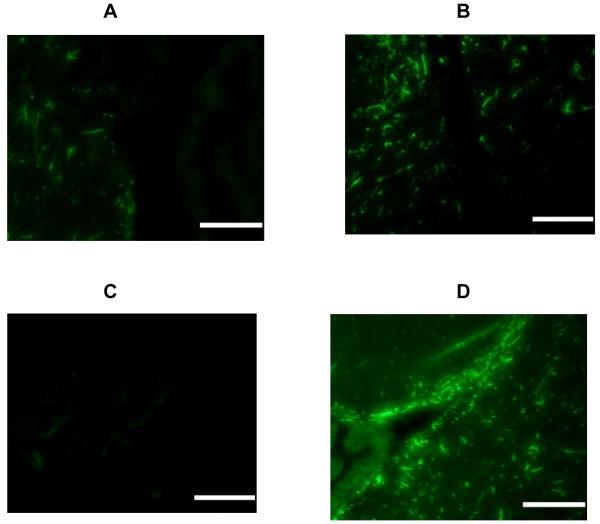
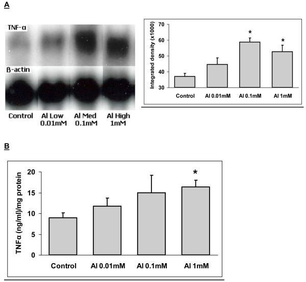
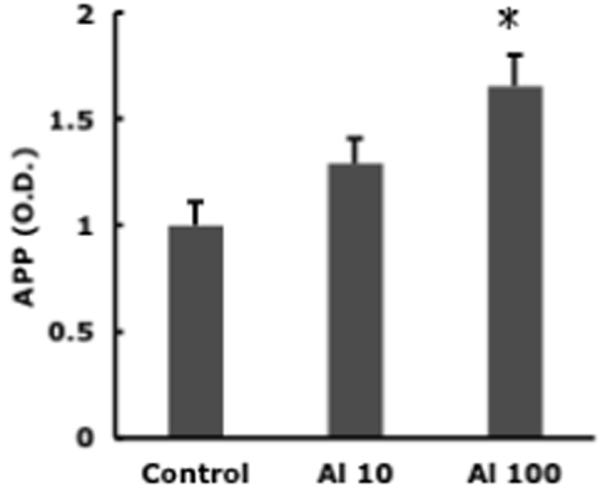
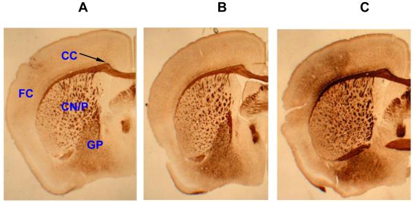
Evidence for the neurotoxicity of extended exposure to low levels of aluminum salts is described using an animal model treated with aluminum at low levels reflecting those found in some water supplies. Emphasis is given to the potential role of aluminum in acceleration and promotion of some indices characteristic of brain aging. These hallmarks include the appearance of excess levels of inflammation in specific brain areas. Aluminum salts can increase levels of glial activation, inflammatory cytokines and amyloid precursor protein within the brain. Both normal brain aging and to a greater extent, Alzheimer's disease are associated with elevated basal levels of markers for inflammation. These are not attributable to obvious exogenous stimuli and may reflect the lifespan history of the organism's immune responses. It is possible that aluminum salts can act as a subtle promoter of such apparently unprovoked responses.
Copyright © 2010 Elsevier Inc. All rights reserved.
Figures
References
-
- Abou-Donia MB. Metals. In: Abou-Donia MB, editor. Neurotoxicology. CRC Press; Boca Raton FL: 1992. pp. 363–93.
-
- Ahuja M, Bishnoi M, Chopra K. Protective effect of minocycline, a semi-synthetic second-generation tetracycline against 3-nitropropionic acid (3-NP)-induced neurotoxicity. Toxicology. 2008;244:111–22. - PubMed
-
- Andrasi E, Pali N, Molnar Z, Kosel S. Brain aluminum, magnesium and phosphorus contents of control and Alzheimer-diseased patients. J Alzheimers Dis. 2005;7:273–84. - PubMed
-
- Atwood CS, Moir RD, Huang X, Scarpa RC, Bacarra NM, Romano DM, Hartshorn MA, Tanzi RE, Bush AI. Dramatic aggregation of Alzheimer abeta by Cu(II) is induced by conditions representing physiological acidosis. J Biol Chem. 1998;273:12817–26. - PubMed
Publication types
MeSH terms
Substances
Grants and funding
LinkOut - more resources
Full Text Sources
Other Literature Sources
