. 2010 Aug;14(8):365-75.
doi: 10.1016/j.tics.2010.05.003. Epub 2010 Jun 16.

Dynamic Network Connectivity: A new form of neuroplasticity

Affiliations

Dynamic Network Connectivity: A new form of neuroplasticity

Amy F T Arnsten et al. Trends Cogn Sci. 2010 Aug.

Abstract

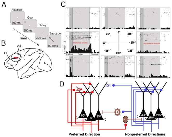
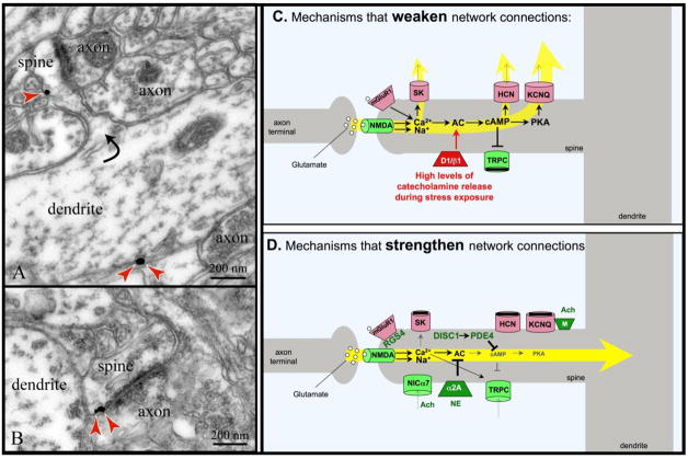
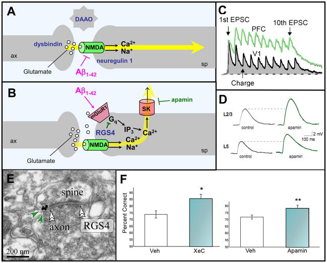
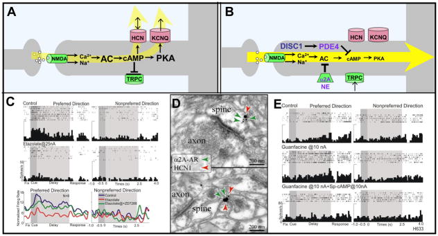
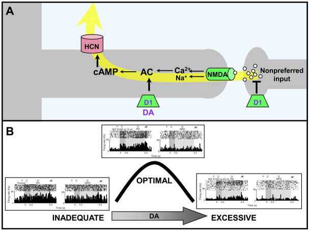
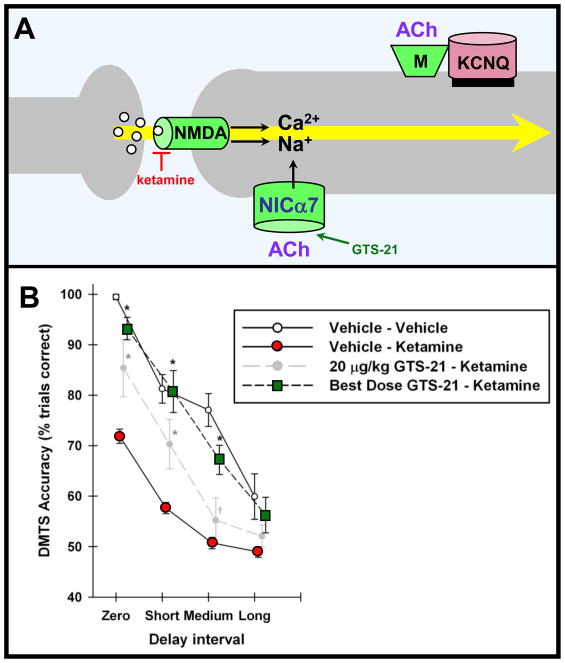
Prefrontal cortical (PFC) working memory functions depend on pyramidal cell networks that interconnect on dendritic spines. Recent research has revealed that the strength of PFC network connections can be rapidly and reversibly increased or decreased by molecular signaling events within slender, elongated spines: a process we term Dynamic Network Connectivity (DNC). This newly discovered form of neuroplasticity provides great flexibility in mental state, but also confers vulnerability and limits mental capacity. A remarkable number of genetic and/or environmental insults to DNC signaling cascades are associated with cognitive disorders such as schizophrenia and age-related cognitive decline. These insults can dysregulate network connections and erode higher cognitive abilities, leading to symptoms such as forgetfulness, susceptibility to interference, and disorganized thought and behavior.


Figures

Box 1 Figure 1
Box 1 Figure 1
The neural circuitry underlying spatial working memory task as envisioned by Goldman-Rakic and colleagues [6]. A. The oculomotor delayed response (ODR) task, a test of spatial working memory. B. The region of the monkey dorsolateral PFC dedicated to spatial working memory. PS=principal sulcus; AS=arcuate sulcus. C. A representative neuron in PFC with spatially tuned firing during the delay period of the ODR task. For details, see [7]. D. The PFC microcircuits subserving spatially tuned firing during the delay period in a spatial working memory task.
Figure 1
Figure 1
A working model of DNC mechanisms. DNC signaling proteins are often found in long, thin spines with narrow spine necks in the superficial layers of monkey dorsolateral PFC. A. Immunoelectron micrograph of HCN channels (red arrows) on a spine neck across from an asymmetric (presumed glutamatergic) synapse, and on the parent dendrite. HCN channels in spines appear to gate synaptic inputs onto the spines, while those on dendrites may regulate excitability (adapted from [7]). B. Immunoelectron micrograph of HCN channels (red arrows) on a spine head next to the post-synaptic density of an asymmetric (presumed glutamatergic) synapse (adapted from [7]). C. Working model of molecular mechanisms that weaken PFC network connectivity. D. Working model of molecular mechanisms that strengthen network connectivity. For both C and D, molecules that strengthen connectivity are shown in green; those that weaken connectivity are shown in red.
Figure 2
Figure 2
A working model of glutamate actions at network synapses in monkey dorsolateral PFC. A. Stimulation of NMDA receptors in the synapse mediates network inputs. B. In contrast, stimulation of perisynaptic mGluR1/5 provides negative feedback via Gq signaling. For both A and B, examples of genetic insults in schizophrenia are shown in navy blue; dysbindin regulates glutamate release; DAAO (D-amino acid oxidase) regulates levels of the NMDA modulator, D-serine, while neuregulin 1 regulates NMDA receptor structure and function at the synapse [79]. The actions of soluble Aβ in AD are shown in magenta; Aβ internalizes NMDA receptors in the presence of α7 nicotinic receptors, and stimulates mGluR1. C. Layer V pyramidal cell connections in rat PFC show larger NMDA currents than those in primary visual cortex (V1) in response to a sequence of 10 stimulating pulses and a recovery pulse. Figure courtesy of W.J. Gao with the permission of PNAS USA [9]. D. The NMDA current in rat PFC is increased in the presence of apamin, a selective blocker of SK channels. Figure courtesy of E.S. Faber [16]. E. RGS4 in PFC spines is typically found perisynaptically (double arrowheads point to the synapse) in monkey PFC, where it is strategically positioned to inhibit mGluR1/5-Gq signaling (adapted from [19]). RGS4 is shown in navy blue in B to highlight its reduced expression in PFC in patients with schizophrenia. RGS4 levels are also reduced in AD. F. Blockade of SK channels in rat PFC with apamin, or of IP3-induced Ca2+ release using xestospongin C (XeC), improves working memory performance compared to vehicle (veh) control infusion. Results represent mean ± SEM percent correct. Adapted from [20].
Figure 3
Figure 3
cAMP signaling weakens PFC network connectivity. A. A working model of cAMP signaling mechanisms that weaken network connectivity. cAMP directly increases the open probability of HCN channels, while indirectly affecting KCNQ2/3 channels via PKA. High levels of cAMP also can reduce the depolarizing TRPC current. B. A working model showing mechanisms that inhibit cAMP signaling and strengthen network connections. NE stimulation of α2A-ARs inhibits cAMP production, while DISC1 activates PDE4s to catabolize cAMP. DISC1 is shown in navy blue to highlight its genetic perturbation in some families with schizophrenia. PDE4, NE and α2A-ARs are shown in purple to emphasize their decline with normal aging. C. High levels of cAMP arising from PDE4 inhibition markedly reduce PFC network firing, which can be reversed by blocking HCN channels with ZD7288 (shown) or blocking KCNQ2/3 channels with XE991 or linopirdine (not shown). Reduced PFC network firing also has been seen following increased cAMP signaling with Sp-cAMPS or yohimbine (not shown). Adapted from [7]. D. Dual immunoelectron microscopy showing α2A-AR/HCN1 channel colocalization in the spine head (top) and the spine neck (bottom) in the superficial layers of the monkey dorsolateral PFC. Adapted from [7]. E. Iontophoretic application of the α2A-AR agonist, guanfacine, increases PFC network firing for the preferred direction, while co-application of the cAMP analog, Sp-cAMPS, reverses this effect. Adapted from [7].
Figure 4
Figure 4
DA stimulation of D1Rs weakens PFC network connections. A. Working model showing D1Rs on a different subset of spines than those containing α2A-ARs; the spines receive network inputs from neurons with dissimilar tuning characteristics (e.g. a neuron with a preferred visuospatial direction of 90° receiving an input from a neuron with a preferred response to 120°). D1R stimulation leads to cAMP generation and the opening of ion channels that shunt nearby synaptic inputs. DA and D1R are shown in purple as they decline with normal aging. B. Stimulation with the D1R agonist, SKF8129, produces an inverted-U dose-response whereby moderate, optimal doses enhance spatial tuning by reducing firing during the delay period only for the memory of nonpreferred directions. In contrast, high doses of D1R stimulation suppress firing for all directions. Adapted from [8]. F=fixation; C=cue; D=delay; R=eye movement response; PD=preferred direction of the neuron; NPD=an example of a nonpreferred direction of the neuron.
Figure 5
Figure 5
ACh strengthens PFC network connectivity. A. A working model showing that ACh may strengthen PFC network connections through direct stimulation of α7 nicotinic receptors on spines [54], and indirectly through stimulation of muscarinic receptors which in turn closes KCNQ2/3 channels. Nicotinic α7 receptors are shown in navy blue due to their genetic alteration in some families with schizophrenia; ACh is shown in purple due to its decline with normal aging. B. Systemic administration of the α7 receptor agonist, GTS-21, to monkeys partially rescues the deficits in working memory induced by the NMDA receptor blocker, ketamine. Figure adapted from [52] with the kind permission of A.V. Terry, Jr..

Similar articles

Cited by

References

    1. Arnsten AFT. Stress signaling pathways that impair prefrontal cortex structure and function. Nature Reviews Neuroscience. 2009;32:267–287. - PMC - PubMed
    1. Moore TL, et al. Executive system dysfunction occurs as early as middle-age in the rhesus monkey. Neurobiol Aging. 2006;27:1484–1493. - PubMed
    1. Weinberger DR, et al. Physiologic dysfunction of dorsolateral prefrontal cortex in schizophrenia. I. Regional cerebral blood flow evidence. Archives General Psychiatry. 1986;43:114–124. - PubMed
    1. Rubia K, et al. Hypofrontality in Attention Deficit Hyperactivity Disorder during higher-order motor control: A study with functional MRI. Am J Psychiatry. 1999;156:891–896. - PubMed
    1. Blumberg HP, et al. A functional magnetic resonance imaging study of bipolar disorder: state- and trait-related dysfunction in ventral prefrontal cortices. Arch Gen Psychiatry. 2003;60:601–609. - PubMed

Publication types