. 2010 Aug 20;285(34):26304-14.
doi: 10.1074/jbc.M110.125039. Epub 2010 Jun 16.

Interaction with polyglutamine aggregates reveals a Q/N-rich domain in TDP-43

Affiliations

Interaction with polyglutamine aggregates reveals a Q/N-rich domain in TDP-43

Rodrigo A Fuentealba et al. J Biol Chem. .

Abstract

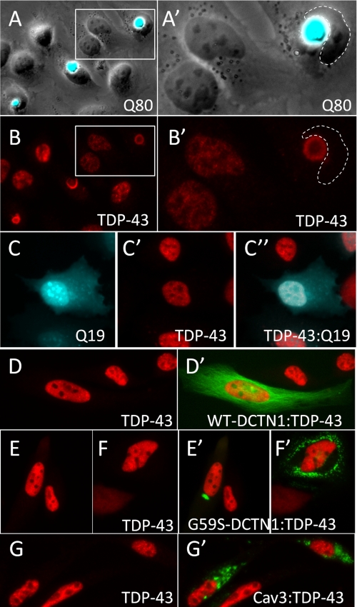
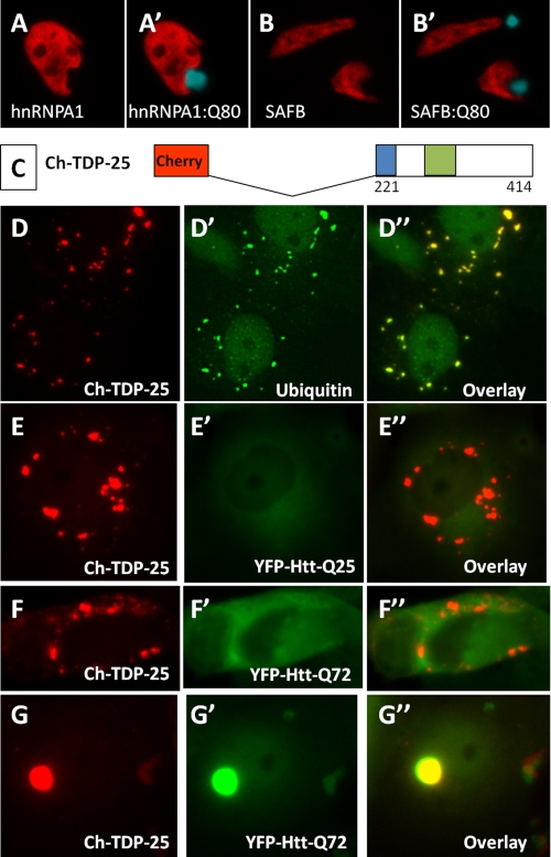
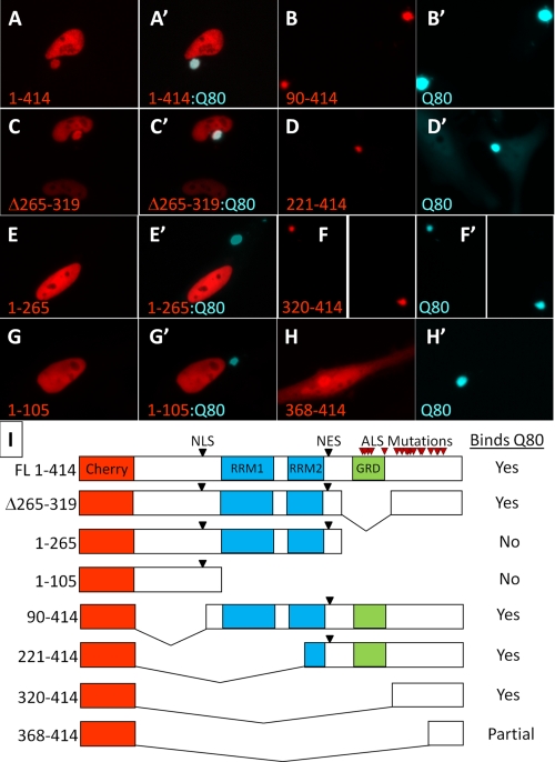
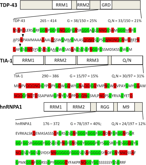
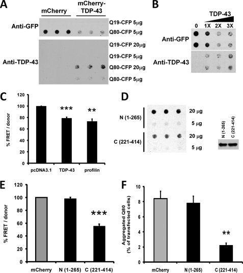
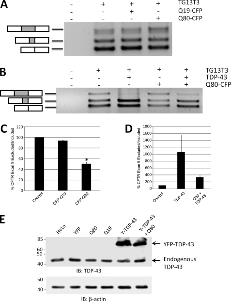
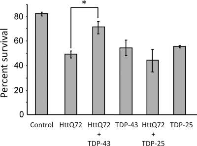
The identification of pathologic TDP-43 aggregates in amyotrophic lateral sclerosis (ALS) and frontotemporal lobar degeneration, followed by the discovery of dominantly inherited point mutations in TDP-43 in familial ALS, have been critical insights into the mechanism of these untreatable neurodegenerative diseases. However, the biochemical basis of TDP-43 aggregation and the mechanism of how mutations in TDP-43 lead to disease remain enigmatic. In efforts to understand how TDP-43 alters its cellular localization in response to proteotoxic stress, we found that TDP-43 is sequestered into polyglutamine aggregates. Furthermore, we found that binding to polyglutamine aggregates requires a previously uncharacterized glutamine/asparagine (Q/N)-rich region in the C-terminal domain of TDP-43. Sequestration into polyglutamine aggregates causes TDP-43 to be cleared from the nucleus and become detergent-insoluble. Finally, we observed that sequestration into polyglutamine aggregates led to loss of TDP-43-mediated splicing in the nucleus and that polyglutamine toxicity could be partially rescued by increasing expression of TDP-43. These data indicate pathologic sequestration into polyglutamine aggregates, and loss of nuclear TDP-43 function may play an unexpected role in polyglutamine disease pathogenesis. Furthermore, as Q/N domains have a strong tendency to self-aggregate and in some cases can function as prions, the identification of a Q/N domain in TDP-43 has important implications for the mechanism of pathologic aggregation of TDP-43 in ALS and other neurodegenerative diseases.


Figures

FIGURE 1.
FIGURE 1.
Cytoplasmic polyglutamine aggregates bind and sequester nuclear TDP-43. A, HeLa cells expressing an expanded polyglutamine construct Q80-CFP developed large cytoplasmic polyglutamine aggregates (A, overlay of phase contrast and CFP fluorescence images). B, immunofluorescence staining to visualize TDP-43 in cells with Q80-CFP aggregates showed that endogenous TDP-43 was completely sequestered into the Q80 aggregate and was absent from the nucleus. A′ and B′ represent higher magnification images of the boxed regions shown in A and B. Cells transfected with Q19-CFP (C) showed normal nuclear localization of endogenous TDP-43 (C′, TDP-43, C″- overlay). d–G, TDP-43 immunostaining in HeLa cells transfected other aggregation prone proteins. Wild-type dynactin-1(WT-DCTN1) fused to GFP (D′) showed normal distribution along microtubules, whereas the G59S-DCTN1 mutant formed discrete focal or multifocal ubiquitinated cytoplasmic aggregates (E′ and F′). G, GFP-Caveolin-3 with the P104L point mutation (Cav3) also formed cytoplasmic ubiquitinated aggregates when transfected into HeLa cells. Unlike Q80 polyglutamine aggregates, cytoplasmic aggregates of G59S-DCTN1 (E′ and F′) and Cav3 (G′) did not induce translocation to the cytosol or sequestration of endogenous TDP-43.
FIGURE 2.
FIGURE 2.
Polyglutamine aggregates do not recruit other nuclear RNA-binding proteins, and cytoplasmic aggregates of TDP-43 C-terminal fragments do not recruit polyglutamine proteins. A and B, immunostaining of endogenous hnRNPA1 and SAFB1 in HeLa cells transfected with Q80-CFP. Unlike TDP-43, the nuclear RNA-binding proteins hnRNPA1 and SAFB did not exit the nucleus or bind to cytoplasmic aggregates of Q80-CFP. C, schematic of 25-kDa C-terminal fragment of TDP-43 fused to mCherry, Ch-TDP-25. d, expression of Ch-TDP-25 in COS7 cells showed numerous punctate cytoplasmic aggregates that stained with an antibody to ubiquitin (D′). Co-expression of Ch-TDP-25 and an N-terminal huntingtin fragment containing a normal (E, HttQ25-YFP) or expanded (F, HttQ72-YFP) polyglutamine stretch showed that cytoplasmic aggregates of the C-terminal domain of TDP-43 were common but did not seed aggregation of polyglutamine-containing proteins. By contrast, cells that spontaneously formed aggregates of HttQ72-YFP showed complete sequestration of Ch-TDP-25 into the aggregate (G).
FIGURE 3.
FIGURE 3.
Polyglutamine aggregate interaction requires amino acids 320–367 in the C-terminal domain of TDP-43. To determine the region of TDP-43 necessary for interaction with polyglutamine aggregates, a series of deletion mutants in TDP-43 fused to mCherry were individually co-transfected into HeLa cells with Q80-CFP, and imaged using fluorescence microscopy. Full-length TDP-43 (A) and deletion of the glycine-rich domain (C) both localized properly to the nucleus and were sequestered into the Q80-CFP aggregate. C-terminal deletions (1–105 and 1–265) retaining the nuclear localization signal (NLS) were localized to the nucleus but did not bind to Q80-CFP aggregates (E and G). N-terminal deletions missing the nuclear localization signal were localized to the cytoplasm (see Fig. 2D); however, in the presence of Q80-CFP aggregates all of the cytoplasmic TDP-43 fragments were incorporated into polyglutamine inclusions (B, D, and F), with the exception of 368–414, which showed only partial colocalization with Q80-CFP (H). The core domain required for binding to polyglutamine aggregates was amino acids 320–367, immediately adjacent to the GRD. I, schematic table of deletion constructs and their binding to Q80-CFP, showing location of the nuclear localization and export signals (NLS and NES), RNA-binding motifs (RRM1 and RRM2), and the location of ALS mutations in TDP-43 (red arrowheads).
FIGURE 4.
FIGURE 4.
Sequence of the C-terminal domains of TDP-43, TIA-1, and hnRNPA1 reveals a Q/N-rich domain in TDP-43. TDP-43 is shown at the top, followed by TIA-1, which contains a well characterized Q/N-rich domain at the C terminus, and hnRNPA1, which contains a canonical glycine-rich domain of the 2×RBD-Gly family, composed of an RGG domain and an M9 nuclear shuttling signal. Glycine (G) residues are highlighted in green and glutamine (Q) and asparagine (N) residues are in red. The overall Q/N content of the C terminus of TDP-43 is 21%. The core region required for the interaction of TDP-43 with polyglutamine aggregates identified in the deletion analysis (flanked by arrowheads; 320–367) shows 31% Q/N content, similar to the Q/N-rich prion-related domain of TIA-1. By contrast, the C-terminal region of hnRNPA1 is significantly more glycine-rich than TDP-43 but has low Q/N content and does not bind polyglutamine aggregates. All but one of the currently described mutations in TDP-43 (underlined) are located in the C-terminal domain.
FIGURE 5.
FIGURE 5.
TDP-43 is sequestered into insoluble polyglutamine aggregates and TDP-43 overexpression inhibits polyglutamine aggregation. A, HeLa cells were transfected with Q19-CFP or Q80-CFP, together with either mCherry (control), or Cherry-TDP-43. Cell lysates (either 5 or 20 μg) were applied to a 0.2-μm pore cellulose acetate filter via vacuum filtration and blotted using antibodies to either GFP (upper panel) or TDP-43 (lower panel) to visualize SDS-insoluble protein aggregates. Q19-CFP, which does not aggregate, passed through the filter, whereas detergent-insoluble aggregates of Q80-CFP were trapped in the filter. Blotting with an anti-TDP-43 antibody showed that in the presence of Q80-CFP, TDP-43 also becomes trapped in the filter, consistent with sequestration into detergent-insoluble Q80-CFP aggregates. Note that TDP-43 transfection decreased the amount of insoluble Q80-CFP retained in the filter (upper panel, right). B, TDP-43 expression decreased Q80-CFP aggregation in a dose-dependent manner. HeLa cells were co-transfected with Q80-CFP and increasing amounts of FLAG-tagged TDP-43 and analyzed using the filter trap assay and anti-GFP antibody. C, TDP-43 decreases the aggregation of expanded huntingtin protein in a FRET assay. HeLa cells were co-transfected with HttQ72-CFP/YFP coding plasmids along with plasmids coding for TDP-43, profilin, or empty vector (pcDNA3.1) and assayed for FRET after 48 h. Calculated FRET was normalized to the donor levels and normalized to pcDNA3.1-transfected cells. Averages from four independent experiments performed in quadruplicate are shown with S.E. as error bars. **, p < 0.01; ***, p < 0.001, paired t test. d, C-terminal (221–414) region of TDP-43 decreases polyglutamine solubility. HeLa cells were co-transfected with Q80-CFP and N-terminal (1–265) or C-terminal (221–414) Cherry-tagged TDP-43 proteins, and filter trap assay was performed with 20 or 5 μg lysates. Right panel, similar level of expression of Q80-CFP in lysates was verified by Western blot. E, TDP-43 fragment containing the Q/N-rich domain decreases aggregation of expanded huntingtin protein. HeLa cells were co-transfected with HttQ72-CFP/YFP along with the indicated mCherry-tagged TDP-43 plasmids and then assayed for FRET after 48 h. Calculated FRET was normalized to the donor levels and represented as % of mCherry-transfected cells. Averages from four independent experiments performed in quadruplicate are shown with S.E. as error bars. ***, p < 0.01, paired t test. F, TDP-43 C-terminal fragment (221–414) reduced inclusion formation, whereas the N-terminal (1–265) fragment did not. HeLa cells were co-transfected with Q80-CFP and either mCherry or the indicated mCherry-tagged TDP-43 constructs. Cells were cultured for 24 h and fixed prior to counting inclusions in at least 2,000 cells from 10 fields per well. The y axis represents the % of transfected cells containing aggregates. **, p < 0.001, t test.
FIGURE 6.
FIGURE 6.
Sequestration of nuclear TDP-43 into polyglutamine aggregates suppresses TDP-43-mediated splicing. To assess whether sequestration of TDP-43 into polyglutamine aggregates had an effect on TDP-43 function, HeLa cells were transfected with a CFTR minigene construct (TG13T3) as a reporter of TDP-43-mediated splicing. In the presence of endogenous TDP-43 levels in HeLa cells, three bands are observed, corresponding to exon 9 inclusion (upper band) or skipping of exon 9 (lower two bands). The middle band is due to the adoption of a cryptic splice acceptor site in exon 9, and the bottom band is from complete exon 9 skipping. A, co-transfection of TG13T3 with Q80-CFP increased exon 9 inclusion, consistent with a loss of basal levels of TDP-43-mediated exon 9 skipping by sequestration into polyQ aggregates. B, overexpression of TDP-43 strongly suppressed exon 9 inclusion (upper band) and enhanced exclusion (lower bands). The alteration in splicing of the CFTR exon 9 minigene induced by Q80-CFP was largely normalized by overexpression of TDP-43. C and d,, mean ratio of exon 9 exclusion/inclusion from three independent experiments, normalized to basal level in HeLa cells (control). *, p < 0.05, paired t test, control versus Q80-CFP. E, immunoblot (IB) of HeLa cell lysates with antibodies to TDP-43, showing that total TDP-43 levels were not affected by transfection with the polyQ-CFP constructs or the YFP-TDP-43 (Y-TDP-43) fusion protein. β-Actin immunoblot is shown below as a loading control.
FIGURE 7.
FIGURE 7.
Increased TDP-43 expression rescues cell death induced by polyglutamine toxicity. COS7 cells were transfected with mCherry alone (Control) or together with the indicated constructs. mCherry positive cells were counted in the same fields at 24 and 72 h after transfection, and percent survival is shown (number of mCherry positive cells at 24 h/number of mCherry positive cells at 72 h) for >200 cells in duplicate wells. HttQ72-induced significant cell death is compared with mCherry alone. However, co-transfection of TDP-43 together with HttQ72 significantly improved cell survival compared with HttQ72 alone (71% versus 49% survival; *, p < 0.05, paired t test). By contrast, expression of the C-terminal fragment of TDP-43(221–414) (TDP-25), which suppresses HttQ72 aggregation but does not localize to the nucleus, did not rescue HttQ72 toxicity. Similar to previous reports, full-length TDP-43 and TDP-25 were also toxic and induced a similar amount of cell death.

References

    1. Neumann M., Sampathu D. M., Kwong L. K., Truax A. C., Micsenyi M. C., Chou T. T., Bruce J., Schuck T., Grossman M., Clark C. M., McCluskey L. F., Miller B. L., Masliah E., Mackenzie I. R., Feldman H., Feiden W., Kretzschmar H. A., Trojanowski J. Q., Lee V. M. (2006) Science 314, 130–133 - PubMed
    1. Gitcho M. A., Baloh R. H., Chakraverty S., Mayo K., Norton J. B., Levitch D., Hatanpaa K. J., White C. L., 3rd, Bigio E. H., Caselli R., Baker M., Al-Lozi M. T., Morris J. C., Pestronk A., Rademakers R., Goate A. M., Cairns N. J. (2008) Ann. Neurol. 63, 535–538 - PMC - PubMed
    1. Yokoseki A., Shiga A., Tan C. F., Tagawa A., Kaneko H., Koyama A., Eguchi H., Tsujino A., Ikeuchi T., Kakita A., Okamoto K., Nishizawa M., Takahashi H., Onodera O. (2008) Ann. Neurol. 63, 538–542 - PubMed
    1. Van Deerlin V. M., Leverenz J. B., Bekris L. M., Bird T. D., Yuan W., Elman L. B., Clay D., Wood E. M., Chen-Plotkin A. S., Martinez-Lage M., Steinbart E., McCluskey L., Grossman M., Neumann M., Wu I. L., Yang W. S., Kalb R., Galasko D. R., Montine T. J., Trojanowski J. Q., Lee V. M., Schellenberg G. D., Yu C. E. (2008) Lancet Neurol. 7, 409–416 - PMC - PubMed
    1. Sreedharan J., Blair I. P., Tripathi V. B., Hu X., Vance C., Rogelj B., Ackerley S., Durnall J. C., Williams K. L., Buratti E., Baralle F., de Belleroche J., Mitchell J. D., Leigh P. N., Al-Chalabi A., Miller C. C., Nicholson G., Shaw C. E. (2008) Science 319, 1668–1672 - PMC - PubMed

Publication types