Comparative Study
. 2010 Jul;133(Pt 7):2123-35.
doi: 10.1093/brain/awq108. Epub 2010 Jun 16.

Kelch-like homologue 9 mutation is associated with an early onset autosomal dominant distal myopathy

Affiliations
Comparative Study

Kelch-like homologue 9 mutation is associated with an early onset autosomal dominant distal myopathy

Sebahattin Cirak et al. Brain. 2010 Jul.

Erratum in

  • Corrigendum.
    [No authors listed] [No authors listed] Brain. 2020 Jun 1;143(6):e52. doi: 10.1093/brain/awaa065. Brain. 2020. PMID: 32163547 Free PMC article. No abstract available.

Abstract

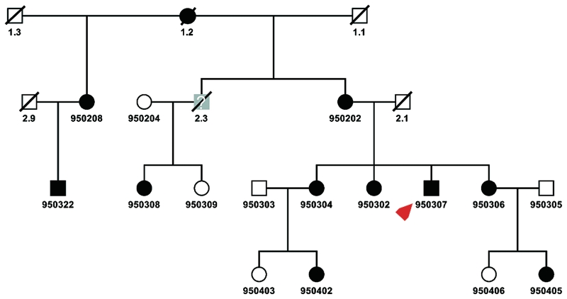
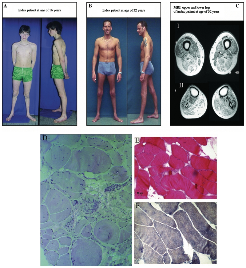
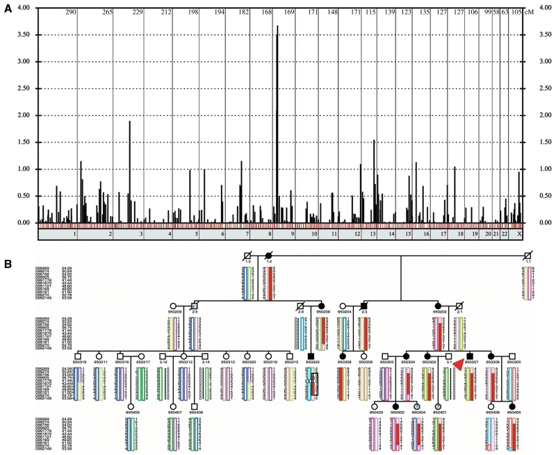
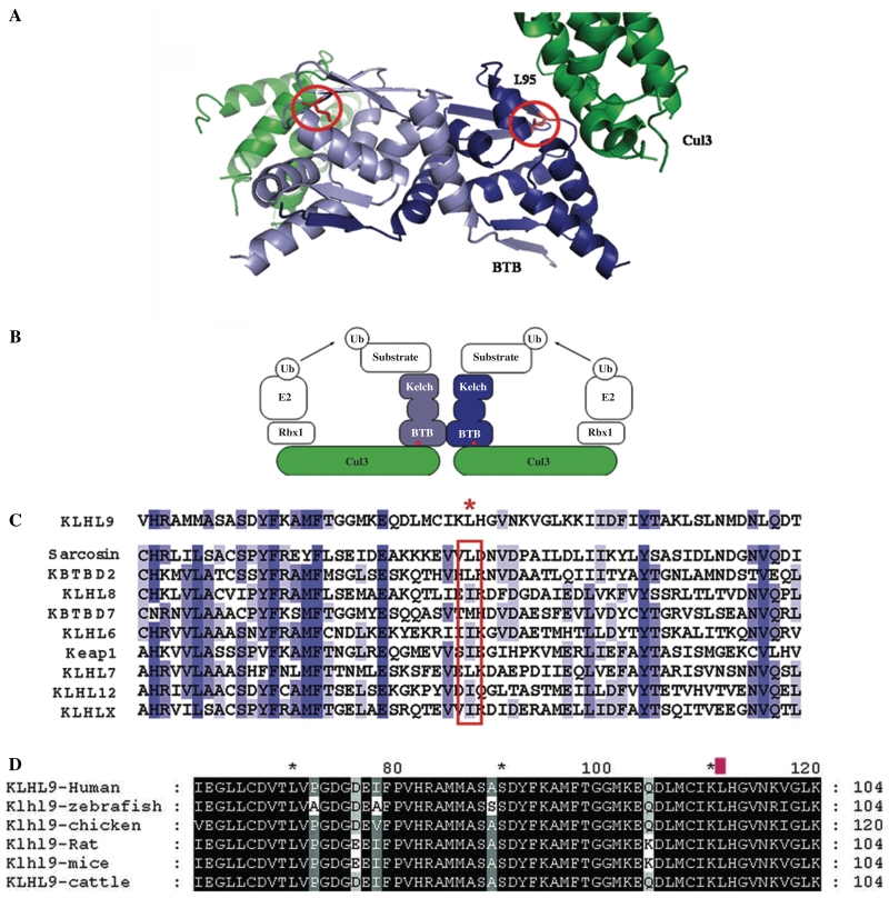
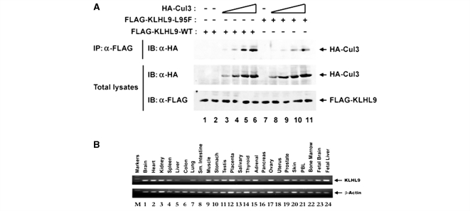
Distal myopathies are a heterogeneous group of disorders characterized by progressive weakness and muscular atrophy, beginning in distal limb muscles and affecting proximal limb muscles at a later stage. We studied a large German kindred with 10 affected members. Weakness and atrophy of the anterior tibial muscles started between the ages of 8 and 16 years, followed by atrophy of intrinsic hand muscles. Progression was slow, and patients retained the ability to walk until the seventh decade. Serum creatinine kinase levels were increased in the range of 150-1400 U/l. Muscle biopsies showed myopathic changes, whereas immunohistochemistry showed normal expression of marker proteins for muscular dystrophies. Patients had reduced sensation with stocking-glove distribution in the distal limbs in later life. Nerve conduction studies revealed no evidence of neuropathy. Genome-wide linkage analysis in this family revealed a new locus for distal myopathy at 9p21.2-p22.3 (multipoint logarithm of the odds ratio=4.21). By positional cloning we found a heterozygous mutation L95F in the Kelch-like homologue 9 gene, encoding a bric-a-brac Kelch protein. Molecular modelling indicated that the mutation may interfere with the interaction of the bric-a-brac domain with Cullin 3. Coimmunoprecipitation experiments confirmed that the mutation reduces association with Cullin 3 in the Kelch-like homologue 9-Cullin 3-E3 ubiquitin ligase complex, which is involved in ubiquitin-dependent protein degradation. We identified a unique form of early onset autosomal dominant distal myopathy which is associated with a Kelch-like homologue 9 mutation and interferes with normal skeletal muscle through a novel pathogenetic mechanism.


Figures

Figure 1
Figure 1
Pedigree. Circles denote females, squares denote males, slashed symbols indicate death, filled symbols indicate the diagnosis of distal myopathy. The index patient is indicated with a red arrow. The symbols filled with grey colour and a question mark indicate that the affection status of these family members could not be identified because of either death or that, at the age of examination, they were considerably younger than the age of onset. All family members with a six-digit identification number were genotyped. This shows the pedigree that was used for the exclusion of distal myopathy loci and the genome scan.
Figure 2
Figure 2
Clinical information from index Patient 950307. (A) Index patient at the age of 16 years, clearly with atrophy of anterior tibial muscles. (B) Index patient at the age of 32 years with the sign of the ‘naked tibia’ due to distal atrophy. (C) Axial T1-weighted spin echo images (Magnetom Symphony, Siemens, FGR: 1.5 Tesla system, repetition time of 377 ms, echo time 20 ms, two acquisitions, 10 mm slice thickness, field of view of 300 × 300 mm) showing the muscle MRI of distal thigh (I) and proximal lower limb (II). In the lower thigh, striking changes are evident in the semimembranosus, biceps femoris and vastus intermedius muscles. Fatty atrophy in the lower leg was pronounced in the tibialis anterior, gastrocnemius and soleus muscles with relative preservation of tibialis posterior and peroneus longus muscles. (D) Trichrom stain of the muscle biopsy from the gastrocnemius muscle at the age of 15 years showing a myopathic pattern with variation fibre size, atrophic fibres, replacement by fat and connective tissue and internal nuclei. Furthermore some peripheral nerves are visualized, which show normal myelinization. (E) Haematoxylin-eosin stain of the vastus lateralis muscle at the age of 32 years confirming the myopathic features and (F) the nicotinamide adenine dinucleotide stain of the same biopsy showing a loss of fibre typing. All images were captured with 20× objective lens.
Figure 3
Figure 3
Whole genome scan and haplotypes of the disease locus at chromosome 9p21.2-p23.3. (A) Genome-wide two point LOD scores are shown, one bar for every genotyped marker. The red bars indicated a maximum LOD score at recombination fraction θ = 0. The blue bars show markers with a positive LOD score with θ > 0, the black bars stand for markers with negative LOD scores. The numbers of the chromosomes are shown under the x-axis, the marker distances are drawn according to the DeCODE Map. (B) This figure shows the haplotypes of the fine mapping using the extended pedigree. Circles denote females, squares denote males, slashed symbols indicate death, and filled symbols indicate the diagnosis of distal myopathy. The symbols filled with grey colour and a question mark indicate that the affection status of these family members could not be identified because of either death or that at the age of examination, they were considerably younger than the age of onset. All family members with a six-digit identification number were genotyped. The haplotypes were reconstructed using the Simwalk2 program. Uninformative or non-reconstructable markers are indicated with a black bar. The order of the microsatellite makers are from p-telomer to centromer according to the DeCODE Map. The disease haplotype is marked with a black square. Note that all affected family members carry the red haplotype. The index case is indicated with a red arrow.
Figure 4
Figure 4
Structural model of the KLHL9–Cul3 complex and alignment of Kelch proteins. (A) The KLHL9 BTB homodimer is shown in light blue/dark blue ribbons and the N-terminal region of Cul3 is shown as two green ribbons. The complex forms a dimer through self-association of the BTB domain, and each BTB domain interacts with the N-terminus of one Cul3 molecule. The two L95 side chains (circled) in the KLHL9 BTB dimer are in the vicinity of two equivalent and independent Cul3-binding interfaces. (B) Schematic representation of the BTB-BACK-Kelch and Cul3 proteins in the context of a full-length, functional ubiquitin ligase complex. The red dots indicate the approximate position of KLHL9 residue L95. (C) Multiple sequence alignment of the BTB domains from a representative subset of the human BTB-BACK-Kelch proteins, including sequences from Val-65 to Thr-125 in KLHL9. The red asterisk denotes the position of L95 in KLHL9. The homologous position in other family members (red box) shows a high propensity for large, aliphatic amino acids. The blue bars indicate the level of conservation from dark blue (highly conserved) to light blue (less conserved). (D) Alignment of several KLHL9 vertebrate proteins. The L95 residue is labelled with a red mark above and shows 100% conservation in vertebrates.
Figure 5
Figure 5
Functional studies and expression. (A) The L95F mutation in KLHL9 reduces association with Cul3. Human embryonic kidney 293T cells were transfected with wild-type cytomegalovirus FLAG-KLHL9 (lanes 1–6) or mutant cytomegalovirus FLAG-L95F KLHL9 (lanes 7–11) and either 0 ng (lanes 1, 2 and 7), 83.3 ng (lanes 3 and 8), 250 ng (lanes 4 and 9), 750 ng (lanes 5 and 10) or 2250 ng of cytomegalovirus HA-Cul3 (lanes 6 and 11) expression vector. Cell lysates were collected at 40 h post-transfection. Equivalent amounts of total protein were immunoprecipitated with α-FLAG antibody and subjected to an α-HA immunoblot (upper panel). Total α-HA (middle panel) and α-FLAG (lower panel) blots were conducted in parallel to confirm the expression of the respective proteins. Our results clearly demonstrate that the p.L95F mutation reduces association with Cul3 (compare lanes 5 to 10 and 6 to 11, for example). (B) KLHL9 is expressed in multiple tissues. To examine the tissue distribution of KLHL9, a human cDNA panel was screened by RT-PCR. PCR products were analysed on a 2% agarose gel. The results were confirmed to be within the linear range by limited dilution of the cDNA products. Quantification of KLHL9 expression is shown in Supplementary Table 1.

Comment in

Similar articles

Cited by

References

    1. Abecasis GR, Cherny SS, Cookson WO, Cardon LR. GRR: graphical representation of relationship errors. Bioinformatics. 2001;17:742–3. - PubMed
    1. Ahlberg G, von Tell D, Borg K, Edstrom L, Anvret M. Genetic linkage of Welander distal myopathy to chromosome 2p13. Ann Neurol. 1999;46:399–404. - PubMed
    1. Ahmad KF, Engel CK, Prive GG. Crystal structure of the BTB domain from PLZF. Proc Natl Acad Sci USA. 1998;95:12123–8. - PMC - PubMed
    1. Aoki M, Arahata K, Brown RH., Jr [Positional cloning of the gene for Miyoshi myopathy and limb-girdle muscular dystrophy] Rinsho Shinkeigaku. 1999;39:1272–5. - PubMed
    1. Bautista J, Rafel E, Castilla JM, Alberca R. Hereditary distal myopathy with onset in early infancy. Observation of a family. J Neurol Sci. 1978;37:149–58. - PubMed

Publication types

MeSH terms