. 2010 Aug 15;21(16):2916-29.
doi: 10.1091/mbc.E10-02-0167. Epub 2010 Jun 16.

Phosphatidic acid induces ligand-independent epidermal growth factor receptor endocytic traffic through PDE4 activation

Affiliations

Phosphatidic acid induces ligand-independent epidermal growth factor receptor endocytic traffic through PDE4 activation

Andrés Norambuena et al. Mol Biol Cell. .

Abstract

Endocytosis modulates EGFR function by compartmentalizing and attenuating or enhancing its ligand-induced signaling. Here we show that it can also control the cell surface versus intracellular distribution of empty/inactive EGFR. Our previous observation that PKA inhibitors induce EGFR internalization prompted us to test phosphatidic acid (PA) generated by phospholipase D (PLD) as an endogenous down-regulator of PKA activity, which activates rolipram-sensitive type 4 phosphodiesterases (PDE4) that degrade cAMP. We found that inhibition of PA hydrolysis by propranolol, in the absence of ligand, provokes internalization of inactive (neither tyrosine-phosphorylated nor ubiquitinated) EGFR, accompanied by a transient increase in PA levels and PDE4s activity. This EGFR internalization is mimicked by PA micelles and is strongly counteracted by PLD2 silencing, rolipram or forskolin treatment, and PKA overexpression. Accelerated EGFR endocytosis seems to be mediated by clathrin-dependent and -independent pathways, leading to receptor accumulation in juxtanuclear recycling endosomes, also due to a decreased recycling. Internalized EGFR can remain intracellular without degradation for several hours or return rapidly to the cell surface upon discontinuation of the stimulus. This novel regulatory mechanism of EGFR, also novel function of signaling PA, can transmodulate receptor accessibility in response to heterologous stimuli.


Figures
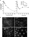
Figure 1.
Figure 1.
PAP inhibition by propranolol induces internalization of EGFR, mimicking the effect of PKA inhibitors. (A) Dose-dependent effect of propranolol on 125I-EGF binding (left). Right panel shows time course with 100 μM propranolol, and the inset shows in detail the kinetics of decrease for the first 10 min (average ± Standard Error of the Mean (SEM) of five independent experiments in triplicate). (B) Indirect immunofluorescence of EGFR. HeLa cells incubated in the absence (Control) or presence of EGF (50 ng/ml) for 10 min, PKA inhibitor PKA-Myr peptide (20 μM), or propranolol (75 μM) for 30 min at 37°C were fixed with 4% paraformaldehyde, permeabilized with 0.2% Triton X-100, and analyzed with HB8506 anti-EGFR antibody. Bar, 10 μm.
Figure 2.
Figure 2.
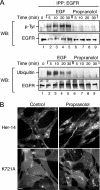
Under propranolol treatment, EGFR is not transactivated and is internalized independently of its tyrosine-kinase activity. (A) Propranolol does not induce tyrosine phosphorylation or ubiquitination of EGFR. HeLa cells were incubated in the absence (lane 1) or presence of either 50 ng/ml EGF (lanes 2–5) or 75 μM propranolol (lanes 6–9) for the indicated time periods at 37°C. EGFR was then immunoprecipitated with mAb-HB8506, and its phosphotyrosine (top) and ubiquitin (bottom) content were assessed by immunoblot with mAb 4G10 anti-phosphotyrosine or anti-ubiquitin mAb P4D1. Bottom, the total mass of EGFR detected after stripping and incubating the blots with polyclonal antibody EGFR984. In contrast with EGF, propranolol does not induce tyrosine phosphorylation or ubiquitination of EGFR. (B) Propranolol induces internalization of K721A kinase-minus EGFR. Indirect immunofluorescence of Her 14 cells expressing either wild type or K721A kinase-minus EGFR shows similar intracellular distribution of both receptors after 30 min of propranolol (75 μM) treatment.
Figure 3.
Figure 3.
PA generated by PLD2 mediates EGFR internalization induced by propranolol. (A) PAP inhibition by propranolol provokes a transient increase of PA levels. HeLa cells were pulse-labeled with [3H]myristic acid (10 μCi) for 16 h and then treated with 75 μM propranolol for 30 min at 37°C. PA levels were resolved by TLC (top) and quantified by scan densitometry. The inhibitory effect of coincubation with 1-butanol (1-OH) is shown for the 20-min time point (top, last lane; bottom, triangle in the graph). (B) RT-PCR for PLD1 and PLD2 mRNAs in HeLa cells transfected twice with the corresponding siRNA for PLD1 or PLD2. The bands were quantified by densitometry (*p < 0.001; two-tailed Student's t test). (C) Counteracting effects of PLD inhibition or silencing. The decrease in 125I-EGF binding induced by 100 μM propranolol (100% effect) is counteracted ∼50% by FIPI, 1-butanol and transfection with siRNA for PLD2 but not PLD1 (*p < 0.001; **p < 0.01). (D) PA directly added in micelles induces EGFR internalization. HeLa cells incubated with PA micelles (400 μg/ml) for 30 min at 37°C show a ∼30% decrease in the levels of 125I-EGF binding (average ± SEM; *p < 0.001). (E) Indirect immunofluorescence of Her14 cells incubated with PA micelles (400 μg/ml) for 30 min at 37°C shows EGFR redistribution from the cell surface to intracellular compartments. Bar, 10 μm.
Figure 4.
Figure 4.
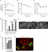
Propranolol-induced EGFR internalization involves the PDE4/cAMP/PKA pathway. (A) Rolipram-sensitive PDE activity increases during propranolol treatment. HeLa cells incubated with 75 μM propranolol show a transient increase in PDE activity that is completely inhibited by 30 μM rolipram (○). (B and C) Propranolol decreases the basal levels of cAMP and PKA. (D) Rolipram inhibits the EGFR internalization induced by propranolol but not by EGF. HeLa cells were incubated with either 75 μM propranolol or 50 ng/ml EGF for 30 min at 37°C, in the presence or absence of 10 or 30 μM rolipram, as indicated. Both concentrations significantly counteracts the effect of propranolol (*p < 0.001). Cells treated with EGF were subjected to acid wash before assessing cell surface 125I-EGF binding at 4°C. (E) Immunofluorescence showing the counteracting effect of rolipram (30 μM). (F) Forskolin counteracts the EGFR internalization induced by propranolol. Cells were preincubated with the indicated concentrations of forskolin for 30 min and then treated with propranolol for another 30 min (*p < 0.001). (G) Overexpression of PKA catalytic subunit abrogates the endocytic effect of propranolol. HeLa cells were transfected with 1 μg PKA-YFP (green) plasmid for 6 h and then incubated with 100 μM propranolol for 30 min and treated for indirect immunofluorescence with anti-EGFR monoclonal antibodies (red). Most of the cells (>97%) expressing PKA-YFP (arrows) do not display internalized EGFR, contrasting with most of the cells not expressing PKA-YFP. Merged image; Bar, 10 μm.
Figure 5.
Figure 5.
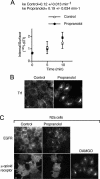
Propranolol does not affect endocytosis of TfR and μ-opioid receptors, but promotes intracellular accumulation of Tf-Alexa 549. (A) Constitutive endocytosis of TfR. HeLa cells were preincubated in serum-free media for 1 h at 37°C and then with 20 ng/ml 125I-diTf for 2 h at 4°C. After eliminating the unbound 125I-diTf, cells were incubated in the presence or absence of 75 μM propranolol for 5–10 min at 37°C. IN/SUR plot shows no difference in the internalization rates (ke). (B) Propranolol increases the accumulation of Tf-Alexa 549 in juxtanuclear endosomes. Cells incubated with 50 μg/ml Tf-Alexa 549 at 37°C for 30 min in the absence or presence of 75 μM and fixed for fluorescence imaging show strong juxtanuclear fluorescence accumulation, suggesting decreased TfR recycling. (C) Propranolol treatment does not cause redistribution of μ-opioid receptors. N2a cells permanently transfected with either EGFR or FLAG-tagged μ-opioid receptors were incubated with either 75 μM propranolol or 10 μM DAMGO (μ-opioid receptor ligand), for 30 min. Bar, 10 μm.
Figure 6.
Figure 6.
Propranolol increases endocytosis (clathrin-dependent and -independent) and inhibits recycling of empty EGFR. (A and B) Endocytosis of cell surface biotinylated EGFR. Cells were biotinylated with cleavable EZ-Link sulfo-NH-SS-biotin at 4°C and then incubated for 5 min at 37°C in the absence (−) or presence (+) of 75 μM propranolol. Glutathione reduction efficiently removes the biotin from the cell surface EGFR (A, and time 0 in B) and reveals a three- to fourfold higher mass of resistant (internalized) EGFR under the effect of propranolol (B), quantified by densitometry (average ± SEM; n + four experiments; *p < 0.05). (C) Top, effect of clathrin silencing with siRNA on EGFR endocytosis. HeLa cells transfected twice, each 24 h, with CHC siRNA show almost complete abrogation (>95%) of clathrin expression, as shown by immunoblot 72 h after the first transfection. Bottom, graph shows the effect of propranolol (100 μM) on 125I-EGF–binding activity in cells transfected with control or clathrin siRNA. Clathrin silencing counteracts only partially (∼50%) this effect, suggesting that a clathrin-independent pathway is also involved (average ± SEM; n + three experiments in triplicate; *p < 0.001). (D) Postendocytic cell surface return of previously internalized biotinylated EGFR. Cells biotinylated with cleavable EZ-Link sulfo-NH-SS-biotin at 4°C were treated for 30 min with propranolol at 37°C, shifted to 4°C, and stripped with glutathione (Initial, internalized pool). A second incubation for another 30 min at 37°C in the absence (−) or presence (+) of 100 μM propranolol followed by glutathione treatment shows three- to fourfold more intracellular EGFR (resistant to glutathione) under the effect of propranolol (graph, average ± SEM; n + four experiments; *p < 0.05). (E) Flow cytometry analysis of cell surface return of previously internalized antibody-EGFR complex. Cells incubated at 4°C with mAb against extracellular EGFR were treated with 100 μM propranolol for 30 min, acid washed at 4°C to remove the remaining cell surface antibodies, and then placed at 37°C for the indicated time periods, in the absence (Control) or presence of propranolol. After incubation with fluorescent secondary antibody, flow cytometry analysis was performed. Progressive cell surface reappearance of EGFR is similar within the first 10–15 min, but afterward ∼90% of EGFR returns to the cell surface in the absence of propranolol (average ± SEM; n + three experiments; *p < 0.05).
Figure 7.
Figure 7.
EGFR internalized during propranolol treatment accumulates in recycling endosomes without degradation. (A) EGFR mass under EGF or propranolol treatment. Immunoblots with polyclonal antibody EGFR984 and densitometric analysis (graph) show an almost unchanged EGFR mass for up to 4 h of propranolol (75 μM) treatment, instead of the progressive decrease (t1/2 ∼60 min) seen during incubation with EGF (50 ng/ml). (B and C) Immunofluorescence colocalization analysis of internalized EGFR. HeLa cells preincubated with 50 μg/ml Tf-Alexa 549 or 1 mM lysotracker at 37°C for 1 h were then treated with either 10 ng/ml EGF (B) or 100 μM propranolol (C) for the indicated time points and then processed for indirect immunofluorescence with anti-EGFR mAb HB8506 (green) and anti-EEA1 polyclonal antibody. Merged digitalized images of representative patterns are shown. Graphs represent the quantitative analysis of EGFR colocalization with each marker. Bar, 10 μm.

Similar articles

Cited by

References

    1. Asp L., et al. Early stages of Golgi vesicle and tubule formation require diacylglycerol. Mol. Biol. Cell. 2009;20:780–790. - PMC - PubMed
    1. Bali P. K., Harris W. R. Site-specific rate constants for iron removal from diferric transferrin by nitrilotris(methylenephosphonic acid) and pyrophosphate. Arch. Biochem. Biophys. 1990;281:251–256. - PubMed
    1. Barbier A. J., Poppleton H. M., Yigzaw Y., Mullenix J. B., Wiepz G. J., Bertics P. J., Patel T. B. Transmodulation of epidermal growth factor receptor function by cyclic AMP-dependent protein kinase. J. Biol. Chem. 1999;274:14067–14073. - PubMed
    1. Baron C. L., Malhotra V. Role of diacylglycerol in PKD recruitment to the TGN and protein transport to the plasma membrane. Science. 2002;295:325–328. - PubMed
    1. Beguinot L., Hanover J. A., Ito S., Richert N. D., Willingham M. C., Pastan I. Phorbol esters induce transient internalization without degradation of unoccupied epidermal growth factor receptors. Proc. Natl. Acad. Sci. USA. 1985;82:2774–2778. - PMC - PubMed

Publication types

MeSH terms