. 2010 Sep;84(17):8650-63.
doi: 10.1128/JVI.00508-10. Epub 2010 Jun 16.

Importin alpha3 interacts with HIV-1 integrase and contributes to HIV-1 nuclear import and replication

Affiliations

Importin alpha3 interacts with HIV-1 integrase and contributes to HIV-1 nuclear import and replication

Zhujun Ao et al. J Virol. 2010 Sep.

Abstract

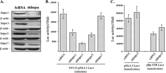
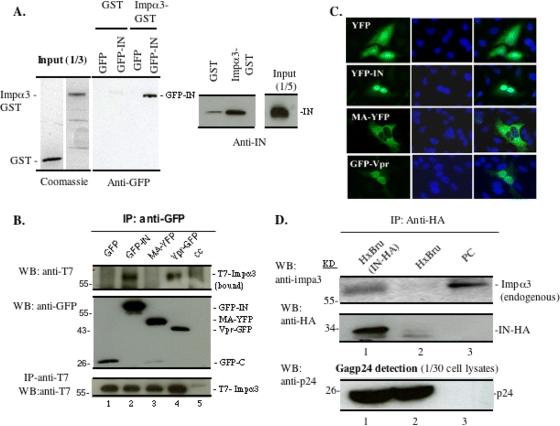
HIV-1 employs the cellular nuclear import machinery to actively transport its preintegration complex (PIC) into the nucleus for integration of the viral DNA. Several viral karyophilic proteins and cellular import factors have been suggested to contribute to HIV-1 PIC nuclear import and replication. However, how HIV interacts with different cellular machineries to ensure efficient nuclear import of its preintegration complex in dividing and nondividing cells is still not fully understood. In this study, we have investigated different importin alpha (Impalpha) family members for their impacts on HIV-1 replication, and we demonstrate that short hairpin RNA (shRNA)-mediated Impalpha3 knockdown (KD) significantly impaired HIV infection in HeLa cells, CD4(+) C8166 T cells, and primary macrophages. Moreover, quantitative real-time PCR analysis revealed that Impalpha3-KD resulted in significantly reduced levels of viral 2-long-terminal repeat (2-LTR) circles but had no effect on HIV reverse transcription. All of these data indicate an important role for Impalpha3 in HIV nuclear import. In an attempt to understand how Impalpha3 participates in HIV nuclear import and replication, we first demonstrated that the HIV-1 karyophilic protein integrase (IN) was able to interact with Impalpha3 both in a 293T cell expression system and in HIV-infected CD4(+) C8166 T cells. Deletion analysis suggested that a region (amino acids [aa] 250 to 270) in the C-terminal domain of IN is involved in this viral-cellular protein interaction. Overall, this study demonstrates for the first time that Impalpha3 is an HIV integrase-interacting cofactor that is required for efficient HIV-1 nuclear import and replication in both dividing and nondividing cells.


Figures

FIG. 1.
FIG. 1.
shRNA-mediated knockdown of Impα1, Impα3, Impα5, and Impα7 in HeLa cells inhibited infection with VSV-G-pseudotyped HIV-1. (A) HeLa cells were transduced with lentiviral vectors harboring Impα1, Impα3, Impα5, or Impα7 shRNA or a scrambled shRNA (ScRNA) and were selected with puromycin (2 μg/ml) for 7 days. Cells were subsequently analyzed for Impα1, Impα3, Impα5, or Impα7 knockdown (KD) by Western blotting using the corresponding antibodies. β-Actin is included as an internal control. The results shown are representative of three different experiments. (B) A total of 0.2 × 10⁶ HeLa cells with KD of Impα1, Impα3, Impα5, or Impα7, or transduced with a vector harboring ScRNA, were challenged by VSV-G-pseudotyped, luciferase (Luc)-expressing HIV-1 (pNL-BruΔBgl/Luc) (10 ng virus-associated p24 antigen). The same amount of cells was collected after 48 h of infection and analyzed for Luc activity. (C) HeLa cells with Impα3-KD or ScRNA were transfected with the pNL-BruΔBgl/Luc provirus or an MMLV vector carrying a luciferase gene plasmid (pBp-STR Luc+). Forty-eight hours later, the same amount of cells was collected and analyzed for Luc activity. Data shown are means and standard errors and are representative of the results for duplicate samples from two independent experiments.
FIG. 2.
FIG. 2.
Effects of Impα1-, Impα3-, and Impα5-KD on the infection of CD4+ C8166 cells with VSV-G-pseudotyped HIV-1. (A) C8166 cells were transduced with lentiviral vectors harboring either Impα1, Impα3, or Impα5 shRNA or a scrambled shRNA and were selected with puromycin (0.5 μg/ml) for 7 days. Cells were subsequently analyzed for Impα1, Impα3, or Impα5 expression by Western blotting using the corresponding antibodies. β-Actin was included as an internal control. The results shown are representative of three different experiments. (B) A WST assay was performed to determine the growth of cell populations with Impα1-, Impα3-, or Impα5-KD, or with ScRNA, at different time points, as indicated. (C) A total of 0.5 × 10⁶ C8166 T cells with Impα1-, Impα3-, or Impα5-KD, or with ScRNA, were challenged with VSV-G-pseudotyped, Luc-expressing HIV-1 (pNL-Bru-Luc+/E) (10 ng virus-associated p24 antigen). After 48 h of infection, equal amounts of cells were collected and were analyzed for Luc activity. (D) C8166 T cells with Impα3-KD or with ScRNA were infected with a VSV-G-pseudotyped MLV vector containing the luc gene, and the Luc activity of infected cells was checked at 48 and 72 h postinfection. Data shown are means and standard errors and are representative of the results for duplicate samples from a typical experiment, which were confirmed in two other independent experiments.
FIG. 3.
FIG. 3.
Impα3 knockdown significantly inhibited the infection of CD4+ C8166 T cells with wild-type HIV-1. (A) Inhibitory effect of Impα3-KD on HIV-1 replication and progression in CD4+ C8166 T cells. A total of 0.5 × 10⁶ Impα3-KD or ScRNA-transduced C8166 cells were infected with HIV-1 pNL4.3-GFP at an MOI of 0.02. At various days postinfection (x axis), the supernatant was collected, and HIV p24gag levels were measured in order to monitor virus replication. (B) After 4 days of infection, the infected (GFP-positive) Impα3-KD and ScRNA-transduced C8166 cells were visualized by fluorescence microscopy (left), and the viral protein p24gag was detected by Western blotting using an anti-p24 antibody (right). (C) Levels of expression of the CD4 receptor on the surfaces of Impα3-KD, ScRNA-transduced, or normal (Mock) C8166 T cells were observed by anti-CD4 staining and flow cytometry analysis.
FIG. 4.
FIG. 4.
Effects of Impα3-KD on HIV reverse transcription, the formation of 2-LTR DNA, and the level of integrated provirus. Impα3-KD and ScRNA-transduced C8166 T cells were infected with HIV-1 pNL4.3-GFP for 2 h, washed twice, and cultured in RPMI medium. At distinct time points after infection (12 and 24 h), DNA was extracted from infected cells, and HIV-1 late reverse transcription products (A), HIV-1 2-LTR circles (B), and the integrated DNA level (C) were analyzed by RQ-PCR using the corresponding primers as described in Materials and Methods. Data shown are means and standard errors and are representative of the results for duplicate samples from a typical experiment, which were confirmed in two other independent experiments.
FIG. 5.
FIG. 5.
Impα3 interacts with HIV-1 integrase (IN) in vitro and in cotransfected 293T cells and HIV-infected C8166 T cells. (A) (Left) Cell lysates from 293T cells expressing GFP or GFP-IN were incubated with either GST alone or a recombinant GST-Impα3 fusion protein. After GST pulldown, the bound proteins were analyzed by Western blotting (WB) with an anti-GFP antibody. (Right) Equal amounts of GST and GST-Impα3 were incubated with purified recombinant HIV-1 IN followed by GST pulldown and were analyzed on an SDS-PAGE gel by Western blotting with rabbit anti-IN antibodies. (B) Impα3 interacts with HIV-1 IN and Vpr in 293T cells. A full-length human T7-Impα3 expression plasmid was cotransfected with a plasmid expressing GFP, GFP-IN, MA-YFP, or Vpr-YFP into 293T cells. (Top) After 48 h of transfection, 90% of the transfected cells were lysed in 0.25% NP-40 buffer and were immunoprecipitated with a rabbit anti-GFP antibody. The immunoprecipitates were resolved using 10% SDS-PAGE, and the bound T7-Impα3 was detected by WB with an anti-T7 antibody. (Center) The presence of GFP-INwt/mut in immunoprecipitates was detected by an anti-GFP antibody. (Bottom) To check protein expression, the remaining 10% of the transfected cells were lysed with 0.5% NP-40, run directly on a 10% SDS-PAGE gel, and probed with anti-T7 antibodies. (C) HeLa cells were transfected with a GFP, GFP-IN, MA-YFP, or Vpr-YFP plasmid. After 48 h, cells were fixed and labeled with an anti-GFP antibody, and the nuclei were stained with 4′,6-diamidino-2-phenylindole (DAPI). Cells were then analyzed by fluorescence microscopy (with a 40× objective lens). (D) Impα3 interacts with HIV-1 IN in virus-infected C8166 T cells. Approximately 10 × 106 C8166 T cells were infected with an HxBru or HxBru-IN-HA virus. (Top) Seventy-two hours after infection, cells were lysed and immunoprecipitated with a rabbit anti-HA antibody, and the bound endogenous Impα3 was detected by Western blotting using rabbit anti-Impα3. The normal C8166 cell lysate was loaded as a positive control (PC). (Center) The immunoprecipitated HA-IN was also detected by WB using a mouse anti-HA antibody. (Bottom) Lysates from the infected C8166 cells were directly loaded onto an SDS-PAGE gel to detect HIV p24gag protein by WB using an anti-p24 antibody.
FIG. 6.
FIG. 6.
Requirement of the C-terminal domain of HIV-1 IN for IN-Impα3 interaction. (A) Plasmids encoding either GFP, GFP-IN, the C-terminal deletion mutant GFP-IN1-212, or the N-terminal deletion mutant GFP-IN50-288 were each cotransfected with the T7-Impα3 expression plasmid into 293T cells, and the interaction of each form of IN with Impα3 was analyzed by anti-GFP immunoprecipitation followed by Western blotting (WB) with an anti-T7 antibody. (Top) Bound T7-Impα3 was detected by IP with an anti-GFP antibody followed by WB using an anti-T7 antibody. (Center) The immunoprecipitated GFP, wild-type GFP-IN, and GFP-IN deletion mutants were detected by WB with an anti-GFP antibody. (Bottom) T7-Impα3 expression was detected with an anti-T7 antibody. (B) Plasmids expressing wild-type GFP-IN or the different IN C-terminal deletion mutants, including GFP-IN1-212, GFP-IN1-250, and GFP-IN1-270, were each cotransfected with a T7-Impα3 expression plasmid into 293T cells, and the Impα3 binding of each IN mutant was analyzed by the co-IP assay as described in Materials and Methods. (Top) Bound T7-Impα3 was detected by IP with anti-GFP followed by WB using anti-T7 antibodies. (Center) The immunoprecipitated GFP, wild-type GFP-IN, and GFP-IN deletion mutants were detected by WB with an anti-GFP antibody. (Bottom) Total T7-Impα3 expression in the transfected cells was examined by Western blotting using an anti-T7 antibody. (C). Intracellular localizations of different GFP-IN mutants. HeLa cells were transfected with plasmids expressing GFP, GFP-IN, GFP-IN1-212, GFP-IN1-250, or GFP-IN1-270. After 48 h, cells were fixed and labeled with an anti-GFP antibody, and the nuclei were stained with 4′,6-diamidino-2-phenylindole (DAPI). Cells were then analyzed by fluorescence microscopy (with a 40× objective lens).
FIG. 7.
FIG. 7.
Inhibitory effect of Impα3-KD on HIV-1 infection in human primary macrophages. (A) MDMs from different donors were transduced with equal amounts of lentiviral vectors carrying ScRNA or Impα3 shRNA. (Top) At day 4 posttransduction, endogenous Impα3 expression in Impα3 shRNA- and ScRNA-treated macrophages was detected by Western blotting using an anti-Impα3 antibody. β-Actin was included as an internal control. (Bottom) Nontransduced (a), ScRNA-transduced (b), and Impα3 shRNA-transduced (c) MDMs were observed under a microscope with a 20× objective lens. (B and C) Impα3 shRNA- or ScRNA-transduced MDMs from donor 1 or donor 2 were infected with VSV-G-pseudotyped pNL-Bru-Luc+/R+ HIV-1. Equal amounts of cells were collected and analyzed for Luc activity at various days postinfection (B) or at day 7 (C). (D) Impα3 shRNA- or ScRNA-transduced MDMs from donor 3 were infected with a VSV-G-pseudotyped R+ or R pNL-Bru-Luc+ virus. After 7 days of infection, equal amounts of cells were collected and analyzed for Luc activity.

Similar articles

Cited by

References

    1. Ao, Z., K. R. Fowke, E. A. Cohen, and X. Yao. 2005. Contribution of the C-terminal tri-lysine regions of human immunodeficiency virus type 1 integrase for efficient reverse transcription and viral DNA nuclear import. Retrovirology 2:62. - PMC - PubMed
    1. Ao, Z., G. Huang, H. Yao, Z. Xu, M. Labine, A. W. Cochrane, and X. Yao. 2007. Interaction of human immunodeficiency virus type 1 integrase with cellular nuclear import receptor importin 7 and its impact on viral replication. J. Biol. Chem. 282:13456-13467. - PubMed
    1. Ao, Z., X. Yao, and E. A. Cohen. 2004. Assessment of the role of the central DNA flap in human immunodeficiency virus type 1 replication by using a single-cycle replication system. J. Virol. 78:3170-3177. - PMC - PubMed
    1. Ao, Z., Z. Yu, L. Wang, Y. Zheng, and X. Yao. 2008. Vpr14-88-Apobec3G fusion protein is efficiently incorporated into Vif-positive HIV-1 particles and inhibits viral infection. PLoS One 3:e1995. - PMC - PubMed
    1. Aratani, S., T. Oishi, H. Fujita, M. Nakazawa, R. Fujii, N. Imamoto, Y. Yoneda, A. Fukamizu, and T. Nakajima. 2006. The nuclear import of RNA helicase A is mediated by importin-α3. Biochem. Biophys. Res. Commun. 340:125-133. - PubMed

Publication types