. 2010 Sep;84(17):8446-59.
doi: 10.1128/JVI.01416-09. Epub 2010 Jun 16.

Coxsackievirus B3 infection activates the unfolded protein response and induces apoptosis through downregulation of p58IPK and activation of CHOP and SREBP1

Affiliations

Coxsackievirus B3 infection activates the unfolded protein response and induces apoptosis through downregulation of p58IPK and activation of CHOP and SREBP1

Huifang M Zhang et al. J Virol. 2010 Sep.

Abstract

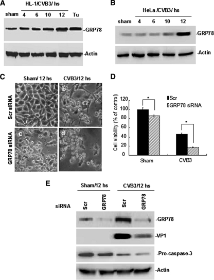
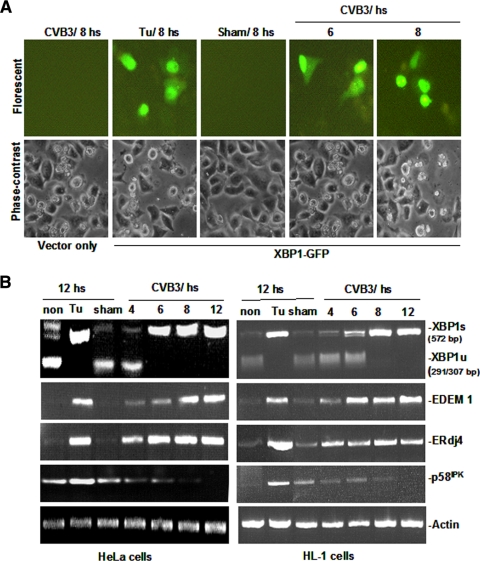
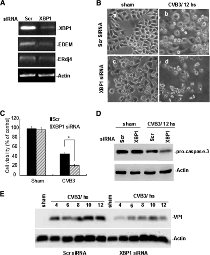
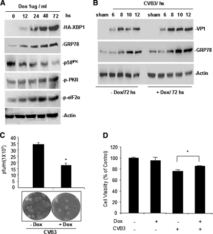
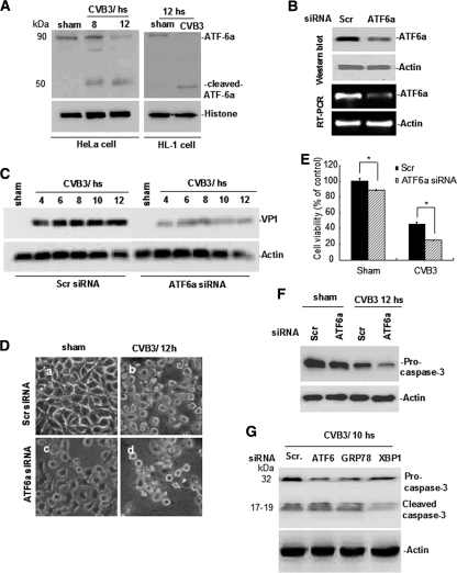
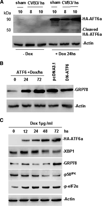
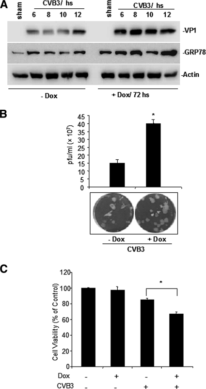
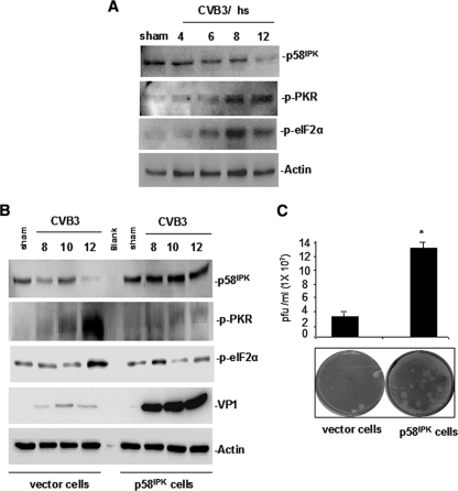
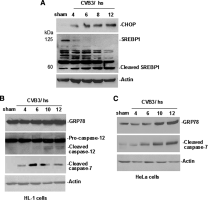
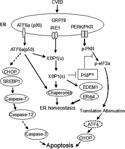
Cardiomyocyte apoptosis is a hallmark of coxsackievirus B3 (CVB3)-induced myocarditis. We used cardiomyocytes and HeLa cells to explore the cellular response to CVB3 infection, with a focus on pathways leading to apoptosis. CVB3 infection triggered endoplasmic reticulum (ER) stress and differentially regulated the three arms of the unfolded protein response (UPR) initiated by the proximal ER stress sensors ATF6a (activating transcription factor 6a), IRE1-XBP1 (X box binding protein 1), and PERK (PKR-like ER protein kinase). Upon CVB3 infection, glucose-regulated protein 78 expression was upregulated, and in turn ATF6a and XBP1 were activated via protein cleavage and mRNA splicing, respectively. UPR activity was further confirmed by the enhanced expression of UPR target genes ERdj4 and EDEM1. Surprisingly, another UPR-associated gene, p58(IPK), which often is upregulated during infections with other types of viruses, was downregulated at both mRNA and protein levels after CVB3 infection. These findings were observed similarly for uninfected Tet-On HeLa cells induced to overexpress ATF6a or XBP1. In exploring potential connections between the three UPR pathways, we found that the ATF6a-induced downregulation of p58(IPK) was associated with the activation of PKR (PERK) and the phosphorylation of eIF2alpha, suggesting that p58(IPK), a negative regulator of PERK and PKR, mediates cross-talk between the ATF6a/IRE1-XBP1 and PERK arms. Finally, we found that CVB3 infection eventually produced the induction of the proapoptoic transcription factor CHOP and the activation of SREBP1 and caspase-12. Taken together, these data suggest that CVB3 infection activates UPR pathways and induces ER stress-mediated apoptosis through the suppression of P58(IPK) and induction/activation of CHOP, SREBP1, and caspase-12.


Figures

FIG. 1.
FIG. 1.
CVB3 infection upregulates GRP78 expression, and the silencing of GRP78 enhances CVB3-induced cell death and reduces VP1 synthesis. Mouse cardiomyocyte HL-1 cells (A) and HeLa cells (B) were infected with CVB3 at an MOI of 100 and 10, respectively. The cell lysates were prepared at the indicated time points pi and subjected to Western blot analysis using the indicated antibodies. HeLa cells treated with tunicamycin (Tu) (2 mg/ml) to induce ER stress were included as a positive control. Actin is detected as a loading control. HeLa cells were transfected with GRP78 siRNA or scrambled (Scr) siRNA and then infected with CVB3. At 12 h pi, cell morphology was observed by phase-contrast microscopy (C), and cell viability was measured by MTS assay and converted to a percentage of a control receiving no siRNA transfection and sham infection (D). Error bars represent means ± SD. P < 0.05. (E) Relative levels of CVB3 VP1 protein and procaspase-3 cleavage were detected by Western blot analysis.
FIG. 2.
FIG. 2.
CVB3 infection induces XBP1 mRNA splicing and alters the expression of its responsive genes. (A) Demonstration of XBP1 splicing by a GFP-based reporter in HeLa cells. Cells were transfected with pHA-XBP1u-GFP and then infected with CVB3, or sham infected with DMEM, or treated only with Tu at a final concentration of 2 mg/ml. Vector-transfected/CVB3-infected cells were included as an additional control. Reporter expression after XBP1 splicing was determined by fluorescent microscopy. (B) RT-PCR analysis. Both HeLa cells and HL-1 cells were infected with CVB3 or sham infected with DMEM. Total cellular RNAs were prepared at the indicated time points pi, and RT-PCRs were performed using specific primers to determine the level of XBP1 splicing and the expression of each indicated target gene. For detecting XBP1 splicing, PCR products were digested with PstI and electrophoresed. The XBP1u (291/307 bp) and XBP1 (572 bp) bands are indicated.
FIG. 3.
FIG. 3.
Silencing of XBP1 enhances CVB3-induced cell death and reduces VP1 synthesis. (A) HeLa cells at 90% confluence were transfected with XBP1 siRNA or scrambled siRNA, and the expression of XBP1 as well as its responsive genes was determined by RT-PCR using primers listed in Table 1. siRNA-transfected HeLa cells were infected with CVB3 or sham infected with DMEM. (B) Cell morphology changes were observed by microscopy at 12 h pi. (C) Cell viability was measured by MTS assay and converted to a percentage of the control as described for Fig. 1. Error bars represent means ± SD. P < 0.05. In addition, procaspase-3 cleavage (D) and CVB3 VP1 production (E) were detected by Western blot analysis using the same amount of cell lysate for each time point. Actin was used as a loading control.
FIG. 4.
FIG. 4.
Overexpression of XBP1 differentially alters target gene expression in the absence or presence of CVB3 infection. (A) Overexpression of XBP1 upregulates GRP78 but downregulates p58IPK in the absence of CVB3 infection. Tet-On/HA-XBP1 HeLa cells were induced (+Dox) or not induced (−Dox) for XBP1 expression. Cell lysates were subjected to Western blot analysis to detect XBP1 and its target genes, GRP78 and p58IPK. (B) Overexpression of XBP1 promotes CVB3 VP1 protein synthesis. Tet-On/HA-XBP1 HeLa cells were left uninduced or were induced with Dox and then infected with CVB3. At the indicated time points pi, cell lysates were prepared for Western blot analyses of CVB3 VP1 and GRP78 proteins. Actin expression was detected in parallel as a loading control. (C) Viral plaque assay. CVB3 titer was determined at 12 h pi. An uninduced sample was included as a control. (D) Cell viability assay. An MTS assay was performed on the cells described above at 12 h pi. The data are presented as percentages of the uninduced/uninfected control. Error bars represent means ± SD. P < 0.05.
FIG. 5.
FIG. 5.
CVB3 infection induces cleavage of ATF6a, and silencing of ATF6a enhances CVB3-caused cell death and reduces VP1 synthesis. Both HL-1 and HeLa cells were cultured and infected with CVB3 as described for Fig. 1. (A) Cell lysates were prepared and subjected to Western blotting to determine the pattern of ATF6a cleavage. Histone detection serves as a loading control. (B) HeLa cells were transfected with ATF6a siRNA or scrambled siRNA (control). The expression of ATF6a was detected by Western blotting and RT-PCR, as indicated. (C) HeLa cells were infected with CVB3 after siRNA transfection. Cell lysates collected at the indicated time points pi were used to detect VP1 production. (D) Cell morphology was observed by phase-contrast microscopy, and (E) cell viability was measured by MTS assay. The data are presented as a percentage of the control (as described for Fig. 1D). Error bars represent means ± SD. P < 0.05. (F) Western blot of cell lysates collected at 12 h pi, detecting procaspase-3 cleavage. (G) The cleavage of caspase-3 was further confirmed on high-percentage gels on which 80 μg of total protein was loaded. Actin detection serves as a loading control.
FIG. 6.
FIG. 6.
Overexpression of ATF6a alters target gene expression in the absence of CVB3 infection. (A) CVB3-induced cleavage of HA-ATF6a. Tet-On/HA-ATF6a cells were left uninduced or were induced with Dox and then infected with CVB3 or sham infected with PBS. At the indicated time points pi, the functional form of ATF6a (p50) was detected by Western blot analysis. Actin expression was used as a loading control. (B) The specific induction of the UPR in Dox-induced Tet-On/HA-ATF6a cells. HeLa cells were transfected with either the plasmid pcDNA3.1-ATF6(171-373), which overexpresses a dominant-negative (DN) ATF6a, or with the pcDNA3.1 vector only (as a control), and GRP78 expression was detected by Western blot analysis at 48 h posttransfection. GRP78 detected in Tet-On/ATF6a cells after Dox induction was included as an additional control. (C) Overexpression of ATF6a upregulates XBP1 and alters UPR-responsive gene expression in the absence of CVB3 infection. Tet-On/HA-ATF6a cells were induced or not induced for ATF6a expression. Cell lysates were subjected to Western blot analysis to detect the expression levels of ATF6a and other UPR-related genes using the indicated antibodies.
FIG. 7.
FIG. 7.
ATF6a overexpression benefits CVB3 replication. (A) Tet-On/HA-ATF6a HeLa cells were induced or not induced for ATF6a expression and then infected with CVB3. At the indicated time points pi, cell lysates were prepared for Western blot analysis of CVB3 VP1 and GRP78. Actin was detected in parallel as a loading control. (B) Viral plaque assay. The CVB3 titer was determined by plaque assay at 12 h pi. A sample of uninduced cells is included as a control. (C) Cell viability assay. HeLa cells were left uninduced or were induced and then infected with CVB3. Cell viability was determined by MTS assay at 12 h pi. The data are presented as percentages of the uninduced, uninfected control. Error bars represent means ± SD. P < 0.05.
FIG. 8.
FIG. 8.
CVB3-induced downregulation of p58IPK activates PKR-mediated phosphorylation of eIF2α, and overexpression of p58IPK increases CVB3 RNA translation. (A) HeLa cells were cultured and infected with CVB3 as described for Fig. 1. Cell lysates were subjected to Western blot analysis to detect p58IPK and the phosphorylation of PKR and e-IF2α. (B) HeLa cells stably transfected to express p58IPK and vector-transfected cells (as a control) were infected with CVB3 at an MOI of 10. Cell lysates were used to detect p58IPK, CVB3 VP1, and phosphorylated PKR and eIF2α at the indicated time point pi. Actin expression is included as a loading control. (C) The results of viral plaque assays measuring viral titers in infected cell samples collected at 12 h pi are shown.
FIG. 9.
FIG. 9.
CVB3 infection induces upregulation of CHOP and activation of SREBP1, caspase-7, and caspase-12. HeLa cells or HL-1 cells were cultured and infected with CVB3 as described in Fig. 1. Cell lysates were subjected to Western blot analysis to detect the induction and activation (cleavage) of proapoptotic transcription factors CHOP and SREBP1, respectively (A), cleavage pattern (indicating activation) of caspases-7 and caspase-12 in HL-1 cells (B), and caspase-7 in HeLa cells (C). Actin was detected in parallel as a loading control.
FIG. 10.
FIG. 10.
Proposed model of CVB3-induced ER stress response. CVB3 infection activates three arms of UPR pathways and induces apoptosis through the suppression of p58IPK and the activation of CHOP and SREBP1. Notably, the activation of the proapoptotic transcription factors CHOP and SREBP1 likely is through ATF6a and not ATF4. Dotted lines with arrows indicate tentative relationships requiring further confirmation.

Similar articles

Cited by

References

    1. Abbate, A., G. Sinagra, R. Bussani, N. N. Hoke, M. Merlo, A. Varma, S. Toldo, F. N. Salloum, G. G. Biondi-Zoccai, G. W. Vetrovec, F. Crea, F. Silvestri, and A. Baldi. 2009. Apoptosis in patients with acute myocarditis. Am. J. Cardiol. 104:995-1000. - PubMed
    1. Andréoletti, L., D. Hober, P. Becquart, S. Belaich, M. C. Copin, V. Lambert, and P. Wattre. 1997. Experimental CVB3-induced chronic myocarditis in two murine strains: evidence of interrelationships between virus replication and myocardial damage in persistent cardiac infection. J. Med. Virol. 52:206-214. - PubMed
    1. Bechill, J., Z. Chen, J. W. Brewer, and S. C. Baker. 2008. Coronavirus infection modulates the unfolded protein response and mediates sustained translational repression. J. Virol. 82:4492-4501. - PMC - PubMed
    1. Benali-Furet, N. L., M. Chami, L. Houel, F. De Giorgi, F. Vernejoul, D. Lagorce, L. Buscail, R. Bartenschlager, F. Ichas, R. Rizzuto, and P. Paterlini-Brechot. 2005. Hepatitis C virus core triggers apoptosis in liver cells by inducing ER stress and ER calcium depletion. Oncogene 24:4921-4933. - PubMed
    1. Boyce, M., and J. Yuan. 2006. Cellular response to endoplasmic reticulum stress: a matter of life or death. Cell Death Differ. 13:363-373. - PubMed

Publication types

MeSH terms

Substances