. 2010 Sep;84(17):8509-19.
doi: 10.1128/JVI.02176-09. Epub 2010 Jun 16.

Interaction of alphaVbeta3 and alphaVbeta6 integrins with human parechovirus 1

Affiliations

Interaction of alphaVbeta3 and alphaVbeta6 integrins with human parechovirus 1

Jani Seitsonen et al. J Virol. 2010 Sep.

Abstract

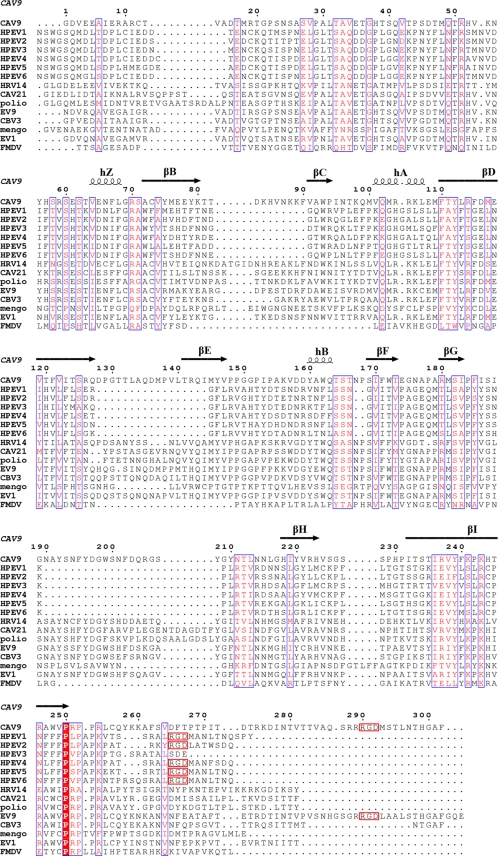
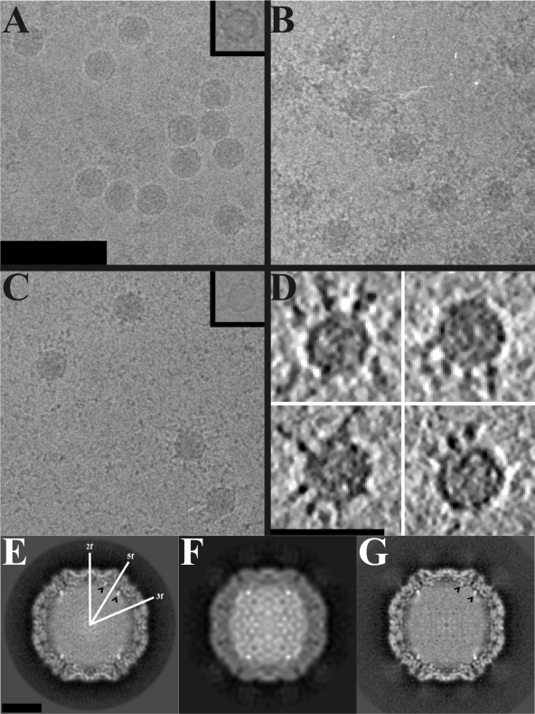
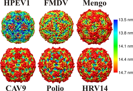
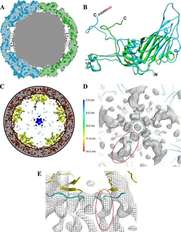
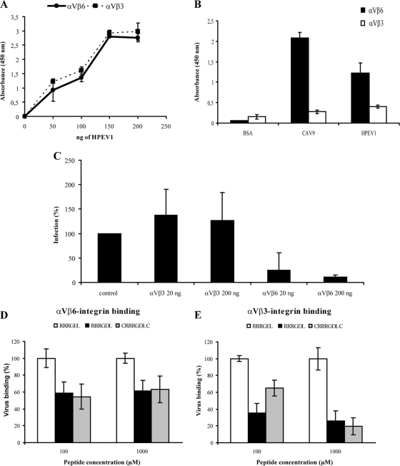
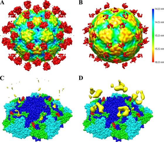
Human parechovirus (HPEV) infections are very common in early childhood and can be severe in neonates. It has been shown that integrins are important for cellular infectivity of HPEV1 through experiments using peptide blocking assays and function-blocking antibodies to alpha(V) integrins. The interaction of HPEV1 with alpha(V) integrins is presumably mediated by a C-terminal RGD motif in the capsid protein VP1. We characterized the binding of integrins alpha(V)beta(3) and alpha(V)beta(6) to HPEV1 by biochemical and structural studies. We showed that although HPEV1 bound efficiently to immobilized integrins, alpha(V)beta(6) bound more efficiently than alpha(V)beta(3) to immobilized HPEV1. Moreover, soluble alpha(V)beta(6), but not alpha(V)beta(3), blocked HPEV1 cellular infectivity, indicating that it is a high-affinity receptor for HPEV1. We also showed that HPEV1 binding to integrins in vitro could be partially blocked by RGD peptides. Using electron cryo-microscopy and image reconstruction, we showed that HPEV1 has the typical T=1 (pseudo T=3) organization of a picornavirus. Complexes of HPEV1 and integrins indicated that both integrin footprints reside between the 5-fold and 3-fold symmetry axes. This result does not match the RGD position predicted from the coxsackievirus A9 X-ray structure but is consistent with the predicted location of this motif in the shorter C terminus found in HPEV1. This first structural characterization of a parechovirus indicates that the differences in receptor binding are due to the amino acid differences in the integrins rather than to significantly different viral footprints.


Figures

FIG. 1.
FIG. 1.
Sequence alignments. Amino acid sequence alignment of the viral coat protein VP1 from different picornaviruses with the CAV9 secondary structure derived from the atomic model displayed above the alignment (34). The columns boxed in blue with red letters signify similarity, and the red column signifies identity. There is limited similarity between HPEV and other picornaviruses. C-terminal RGD motifs are boxed in red.
FIG. 2.
FIG. 2.
Electron cryo-microscopy of HPEV1 and HPEV1 complexed with integrins. (A) Micrograph of HPEV1 at 2.8-μm underfocus. (B) Micrograph of HPEV1 complexed with integrin αVβ3 at 2.5-μm underfocus. (C) Micrograph of HPEV1 complexed with integrin αVβ6 at 3.1-μm underfocus. Scale bar, 100 nm (A; also for B and C). An empty particle image is inset in panels A and C. (D) Slices (0.76 nm thick) through tomographic reconstructions of four representative αVβ6-labeled HPEV1 particles. Scale bar, 50 nm. The integrin can be seen bound to the particles in panels B and C, but they are clearest in the tomographic data. (E) Central section of the HPEV1 reconstruction. Twofold, 3-fold, and 5-fold axes of symmetry are indicated. (F) Central section of the HPEV1 complexed with integrin αVβ3 reconstruction. (G) Central section of the HPEV1 complexed with integrin αVβ6 reconstruction. Scale bar, 10 nm (E; also for F and G). The black arrowheads in panels E and G indicate strong fingers of density near a 5-fold axis of symmetry.
FIG. 3.
FIG. 3.
Comparison of the icosahedrally symmetric 0.85-nm-resolution HPEV1 reconstruction with the atomic models of other picornaviruses. HPEV1, FMDV (Protein Data Bank [PDB] code 1bbt), mengovirus (PDB 2mev), CAV9 (PDB 1d4m), poliovirus (1hxs), and HRV14 (4rhv). The X-ray models have been filtered to 0.85-nm resolution, and all models are rendered as isosurface representations at 2 standard deviations above the mean, viewed down a 2-fold axis of symmetry. This comparison demonstrates the diversity of picornavirus surface features as well as the unique features of HPEV1. The models have been colored using radial-depth cueing in CHIMERA (bar, 13.5- to 14.7-nm radius) (60).
FIG. 4.
FIG. 4.
Capsid protein RNA contact. (A) Central section of the 0.85-nm-resolution HPEV1 reconstruction (gray; transparent) with a CAV9 X-ray structure (cyan) fitted on the left-hand side and an FMDV X-ray structure (green) fitted in on the right-hand side of the cryo-EM reconstruction. X-ray structures are presented as sphere models. The CAV9 model protrudes from the HPEV1 density at the 5-folds due to the long surface loops in the β-barrel of CAV9 VP1, whereas FMDV offers a better fit at the 5-fold due to shorter loops. (B) Comparison of CAV9 VP1 (blue) and FMDV VP1 (green) presented as a ribbon. The C and N termini of both CAV9 VP1 and FMDV VP1 are indicated. The loop containing the RGD motif in FMDV is not resolved in the X-ray model, but the nearest three amino acids resolved on both sides of the loop are indicated (red). The extended loops of CAV9 compared to FMDV are also evident in this panel. (C) Slabbed section of the 0.85-nm-resolution HPEV1 reconstruction rendered at 3 standard deviations above the mean, viewed along a 5-fold axis of symmetry and depth queued to reveal the five high-density finger-like protrusions (black arrow) inside the capsid under each vertex. The position of the capsid layer is indicated with a gray ring. (D) A section showing a single set of fingers viewed from the inside the capsid. HPEV1 density is shown as a gray mesh with the superimposed atomic models of CAV9 VP1 (cyan) and VP4 (yellow) in ribbon. This view illustrates how the N termini of CAV9 VP1 and VP4 fit the finger-like densities. (E) The view from panel D tilted 90° and cropped for clarity. This view shows how the finger-like density seen in the HPEV1 reconstruction extends even farther into the RNA than in the atomic models of CAV9 VP1 and VP4. One of the finger-like densities is indicated (circled in red) in panels D and E.
FIG. 5.
FIG. 5.
Integrin binding assays. (A) HPEV1 in vitro binding assay to immobilized integrins (avidity assay). Integrin αVβ3 and αVβ6 (300 ng) were passively immobilized onto microtiter wells, and 0, 50, 100, 150 or 200 ng of HPEV1 was overlaid and incubated for 2 h before detection by virus-specific antibody followed by HRP-conjugated secondary antibody. Wells were incubated with TMB (H2O2 substrate), the reaction was stopped by adding 100 μl of 0.45 M H2SO4, and the absorbance was read at the OD450. Range of two parallel experiments is shown. The experiment was repeated three times with identical results. (B) Binding of integrins to immobilized HPEV1 (affinity assay). A fixed amount (200 ng) of HPEV1 was immobilized onto wells, and binding of 300 ng of integrin αVβ3 and αVβ6 was determined by specific antibodies. Similar amounts of CAV9 (33) and BSA were used as positive and negative controls, respectively. Average values of two independent experiments are shown. (C) Plaque reduction assay with soluble integrins. A total of 1,000 PFU of HPEV1 was incubated with 20 ng or 200 ng of integrins αVβ3 and αVβ6 for 1 h prior to cell infection. Plaques were counted 2 to 4 days postinfection. The infectivity was compared to that of the control (virus incubated without integrins). Data are the average of two independent experiments. (D and E) In vitro peptide blocking assay. Wells were coated with 200 ng of integrins αVβ3 and αVβ6 and preincubated with 100 and 1,000 μM concentrations of peptide RRRGDL, CRRRGDLC, or RRRGEL (as a negative control; 100% virus binding) before virus was added. Virus binding was detected by virus-specific antiserum followed by secondary anti-mouse/rabbit HRP conjugate. Virus binding was calculated by adjusting absorbance values of the control to 100%. The standard deviations of two independent experiments with five measuring points are shown.
FIG. 6.
FIG. 6.
HPEV1-integrin interactions. (A) An isosurface representation of the 1-nm-resolution reconstruction of HPEV1 complexed with integrin αVβ6 at 0.15 standard deviations above the mean density viewed down a 2-fold axis of symmetry and depth queued. (B) An isosurface representation of the 1.5-nm-resolution reconstruction of HPEV1 complexed with integrin αVβ3 at the mean density viewed down a 2-fold axis of symmetry and depth queued. The color key applies to panels A and B. (C) An isosurface representation of just the integrin αVβ6 density at 0.44 standard deviations above the mean in yellow, displayed on top of the CAV9 X-ray model. Residues PTP of CAV9 VP1 are covered by the integrin density. (D) An isosurface representation of just the integrin αVβ3 density (yellow) displayed on top of the CAV9 X-ray model. The reconstruction of HPEV1 complexed with integrin αVβ3 is rendered at the mean to reveal the weak integrin density. The atomic models in panels C and D are shown as a sphere presentation of five copies of VP1, VP2, and VP3. VP1 is shown in dark blue, VP2 is in cyan, and VP3 is in green. The four C-terminal amino acids (VTTV) of the CAV9 VP1 atomic model are shown in red to illustrate the likely position of the RGD loop in CAV9.

Similar articles

Cited by

References

    1. Abed, Y., and G. Boivin. 2005. Molecular characterization of a Canadian human parechovirus (HPeV)-3 isolate and its relationship to other HPeVs. J. Med. Virol. 77:566-570. - PubMed
    1. Abraham, G., and R. J. Colonno. 1984. Many rhinovirus serotypes share the same cellular receptor. J. Virol. 51:340-345. - PMC - PubMed
    1. Acharya, R., E. Fry, D. Stuart, G. Fox, D. Rowlands, and F. Brown. 1989. The three-dimensional structure of foot-and-mouth disease virus at 2.9 Å resolution. Nature 337:709-716. - PubMed
    1. Al-Sunaidi, M., C. H. Williams, P. J. Hughes, D. P. Schnurr, and G. Stanway. 2007. Analysis of a new human parechovirus allows the definition of parechovirus types and the identification of RNA structural domains. J. Virol. 81:1013-1021. - PMC - PubMed
    1. Arita, M., S. Koike, J. Aoki, H. Horie, and A. Nomoto. 1998. Interaction of poliovirus with its purified receptor and conformational alteration in the virion. J. Virol. 72:3578-3586. - PMC - PubMed

Publication types

MeSH terms

LinkOut - more resources