. 2010 Sep;84(17):8470-83.
doi: 10.1128/JVI.00176-10. Epub 2010 Jun 16.

Tick-borne encephalitis virus delays interferon induction and hides its double-stranded RNA in intracellular membrane vesicles

Affiliations

Tick-borne encephalitis virus delays interferon induction and hides its double-stranded RNA in intracellular membrane vesicles

Anna K Overby et al. J Virol. 2010 Sep.

Abstract

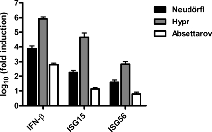
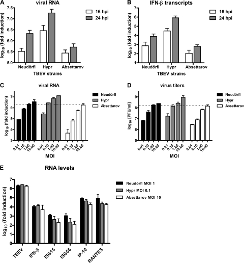
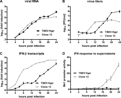
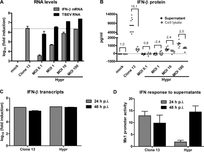
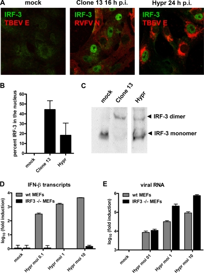
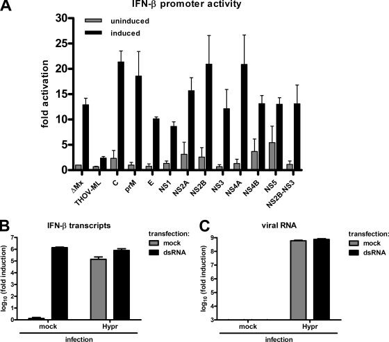
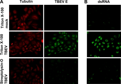
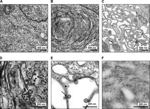
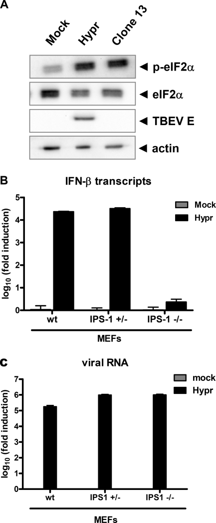
Tick-borne encephalitis virus (TBEV) (family Flaviviridae, genus Flavivirus) accounts for approximately 10,000 annual cases of severe encephalitis in Europe and Asia. Here, we investigated the induction of the antiviral type I interferons (IFNs) (alpha/beta IFN [IFN-alpha/beta]) by TBEV. Using strains Neudörfl, Hypr, and Absettarov, we demonstrate that levels of IFN-beta transcripts and viral RNA are strictly correlated. Moreover, IFN induction by TBEV was dependent on the transcription factor IFN regulatory factor 3 (IRF-3). However, even strain Hypr, which displayed the strongest IFN-inducing activity and the highest RNA levels, substantially delayed the activation of IRF-3. As a consequence, TBEV can keep the level of IFN transcripts below the threshold value that would permit the release of IFN by the cell. Only after 24 h of infection have cells accumulated sufficient IFN transcripts to produce detectable amounts of secreted IFNs. The delay in IFN induction appears not to be caused by a specific viral protein, since the individual expressions of TBEV C, E, NS2A, NS2B, NS3, NS4A, NS4B, NS5, and NS2B-NS3, as well as TBEV infection itself, had no apparent influence on specific IFN-beta induction. We noted, however, that viral double-stranded RNA (dsRNA), an important trigger of the IFN response, is immunodetectable only inside intracellular membrane compartments. Nonetheless, the dependency of IFN induction on IFN promoter stimulator 1 (IPS-1) as well as the phosphorylation of the alpha subunit of eukaryotic initiation factor 2 (eIF2alpha) suggest the cytoplasmic exposure of some viral dsRNA late in infection. Using ultrathin-section electron microscopy, we demonstrate that, similar to other flaviviruses, TBEV rearranges intracellular membranes. Virus particles and membrane-connected vesicles (which most likely represent sites of virus RNA synthesis) were observed inside the endoplasmic reticulum. Thus, apparently, TBEV rearranges internal cell membranes to provide a compartment for its dsRNA, which is largely inaccessible for detection by cytoplasmic pathogen receptors. This delays the onset of IFN induction sufficiently to give progeny particle production a head start of approximately 24 h.


Figures

FIG. 1.
FIG. 1.
Induction of IFN and ISGs. A549 cells were infected with TBEV strains Neudörfl, Hypr, and Absettarov at an MOI 1, and total cell RNA was extracted at 24 h p.i. Levels of IFN-β, ISG15, and ISG56 mRNAs were measured by real-time RT-PCR analysis, normalized to the cellular γ-actin mRNA, and set in relation to mRNA levels of mock-infected cells. Mean values and standard deviations from three independent experiments are shown.
FIG. 2.
FIG. 2.
TBEV strains differ in replication speeds, endpoint titers, and IFN induction capabilities. (A and B) A549 cells were infected with TBEV strains Neudörfl, Hypr, and Absettarov at an MOI of 1, and cellular RNA was extracted either at 16 h p.i. or at 24 h p.i. Viral RNA (A) and IFN-β mRNA levels (B) were quantified by real-time RT-PCR analysis as indicated in the legend of Fig. 1. (C and D) Multiplication of virus strains is dependent on the input MOI. A549 cells were infected with the different TBEV strains by using MOIs varying in 10-fold steps from 0.01 to 10. Total cell RNA and cell culture supernatants were harvested at 24 h p.i., and viral RNA levels (C) and titers (D) were determined by real-time RT-PCR and plaque assays, respectively. (E) Levels of viral RNA and innate immunity genes after infection with TBEV strains using normalized input MOIs. Total RNA preparations from the cells infected for 24 h with the different TBEV strains at MOIs of 1 (Neudörfl), 0.1 (Hypr), and 10 (Absettarov) (see C and D) were used to measure levels of viral RNA and mRNAs for IFN-β, ISG15, ISG56, IP-10, and RANTES by real-time RT-PCR. In all cases, mean values and standard deviations from three independent experiments are shown.
FIG. 3.
FIG. 3.
Time course of viral multiplication, IFN transcription, and IFN production. A549 cells were infected with TBEV strain Hypr and the IFN-inducing control virus clone 13 at an MOI of 1. Total cell RNA and cell culture supernatants were collected at different time points p.i. (A) Quantification of intracellular levels of virus RNA determined by real-time RT-PCR analysis. (B) Viral titers in cell culture supernatants determined by plaque assays. (C) Levels of IFN-β mRNA determined by real-time RT-PCR analysis. (D) Presence of type I IFN in the supernatants of infected cells measured using a 293T cell line carrying the firefly luciferase gene under the control of the IFN-responsive Mx1 promoter (Mx1-luc reporter cells). Mean values and standard deviations from three independent experiments are shown.
FIG. 4.
FIG. 4.
Correlation between transcription, translation, and secretion of IFN. (A and B) Search for comparable IFN induction levels by clone 13 and TBEV. A549 cells were infected for 24 h either with clone 13 at an MOI of 5 or with TBEV strain Hypr at increasing MOIs. (A) Total RNA was extracted from cells, and levels of IFN-β mRNA and clone 13 and TBEV RNAs were determined by real-time RT-PCR analysis. (B) In parallel dishes, supernatants were harvested, and cellular proteins were extracted to measure extracellular (black dots) and intracellular (gray diamonds) IFN-β by ELISA. (C and D) Release of IFN by infected cells. A549 cells were infected with clone 13 (MOI of 5) or TBEV strain Hypr (MOI of 100). (C) Cellular RNA was extracted at 24 h p.i. or 48 h p.i., and levels of IFN-β mRNA were determined by real-time RT-PCR analysis. (D) Either supernatants were harvested at 24 h p.i. or medium was exchanged at 24 h p.i. and harvested at 48 h p.i. IFN levels were measured by using 293T Mx1-luc cells as described in the legend of Fig. 3. Mean values and standard deviations from three independent experiments are shown.
FIG. 5.
FIG. 5.
Involvement of IRF-3. (A) Immunofluorescence analysis. Vero B4 cells were infected at an MOI of 1 with clone 13 or TBEV strain Hypr. At 16 h (clone 13) or 24 h (TBEV) p.i., cells were fixed and immunostained using antisera specific for IRF-3 and viral N antigens, respectively. (B) Percentage of infected cells with nuclear IRF-3 calculated from three independent experiments (100 to 200 cells counted in each experiment), performed as described above (A). (C) Homodimerization assay. Extracts from A549 cells infected with either clone 13 for 16 h or TBEV for 24 h were subjected to nondenaturing gel electrophoresis followed by immunoblotting to detect IRF-3. (D and E) Dependence of IFN induction on IRF-3. wt MEFs (gray bars) and IRF-3 knockout MEFs (black bars) were infected with TBEV at different MOIs, and cellular RNA was extracted at 24 h p.i. Levels of IFN-β transcripts (D) and viral RNA (E) were determined by real-time RT-PCR. Mean values and standard deviations from three independent experiments are shown.
FIG. 6.
FIG. 6.
Screen of TBEV gene products for inhibition of the IFN-β promoter. (A) Reporter assay for testing overexpressed TBEV gene products. A549 cells were transfected with an FF-Luc construct under the control of the IFN-β promoter and a Ren-Luc construct carrying the constitutively active SV40 promoter. In addition, cDNA plasmids expressing individual TBEV genes or a positive (THOV-ML) or a negative (ΔMx) control were transfected. At 24 h posttransfection, cells were infected with clone 13 (MOI of 1) to stimulate the IFN-β promoter. After a further incubation period of 16 h, cells were lysed, and reporter activities were determined. Specific IFN-β promoter activity was determined by normalizing FF-Luc to Ren-Luc activities and setting the mock-infected uninduced ΔMx control as 1. Unstimulated and stimulated values are depicted as gray bars and black bars, respectively. Mean values and standard deviations from three independent experiments are shown. (B and C) Real-time RT-PCR assay of TBEV-infected cells induced with dsRNA. A549 cells were infected with TBEV strain Hypr for 16 h at an MOI of 5 and then transfected with poly(I:C) (dsRNA) for another 6 h before total RNA extraction. Mock infection and mock transfections were performed in parallel as controls. IFN-β mRNA levels (B) and TBEV RNA levels (C) were quantified by real-time RT-PCR analysis. Mean values and standard deviations from three independent experiments are shown.
FIG. 7.
FIG. 7.
Subcellular localization of viral dsRNA. Cells were infected with TBEV (MOI of 1) or left uninfected (mock) and fixed 24 h later with 3% paraformaldehyde. Fixed cells were treated either with Triton X-100, which permeabilizes all cellular membranes, or with SL-O, which selectively permeabilizes the plasma membrane. (A) Immunodetection of tubulin (located in the cytoplasm) and the TBEV E protein (located inside the ER). (B) Immunodetection of dsRNA in cells treated the same way as described above (A).
FIG. 8.
FIG. 8.
Ultrathin-section EM of infected cells. 293T cells were infected with TBEV strain Hypr (MOI of 5) for 24 h before fixation and processing for regular transmission EM (A to E) or immuno-EM (F). (A) Transmission EM of mock-infected cells. (B) Transmission EM of TBEV-infected cells. (C) Transmission EM of TBEV-infected cells treated with BFA at 12 h p.i. (D and E) Close-up EM pictures of untreated and BFA-treated cells that were infected with TBEV. (F) Close-up immuno-EM picture of cells infected with TBEV. Immunogold staining was performed by using anti-dsRNA mouse monoclonal J2 as the primary antibody and goat anti-mouse IgG/IgM conjugated with 10-nm colloidal gold as the secondary antibody. Arrowheads indicate virus particles, lined arrows indicate virus-induced membrane vesicles, and the boxed arrow indicates immunogold-labeled dsRNA.
FIG. 9.
FIG. 9.
Cytoplasmic detection of viral RNA late in infection. (A) eIF2α activation. A549 cells were infected with TBEV strain Hypr for 24 h and then assayed by Western blot analysis using antisera recognizing phosphorylated eIF2α (p-eIF2α), eIF2α, TBEV E, or actin as a control. (B and C) Real-time RT-PCR assay. MEF cells expressing or lacking one or both genomic copies of the IPS-1 gene were infected with Hypr at an MOI of 1, and total cell RNA was extracted at 24 h p.i. Mock infections were performed in parallel as controls. IFN-β mRNA levels (B) and TBEV RNA levels (C) were quantified by real-time RT-PCR analysis. Mean values and standard deviations from three independent experiments are shown.

Similar articles

Cited by

References

    1. Ahlquist, P. 2006. Parallels among positive-strand RNA viruses, reverse-transcribing viruses and double-stranded RNA viruses. Nat. Rev. Microbiol. 4:371-382. - PMC - PubMed
    1. Ashour, J., M. Laurent-Rolle, P. Y. Shi, and A. Garcia-Sastre. 2009. NS5 of dengue virus mediates STAT2 binding and degradation. J. Virol. 83:5408-5418. - PMC - PubMed
    1. Best, S. M., K. L. Morris, J. G. Shannon, S. J. Robertson, D. N. Mitzel, G. S. Park, E. Boer, J. B. Wolfinbarger, and M. E. Bloom. 2005. Inhibition of interferon-stimulated JAK-STAT signaling by a tick-borne flavivirus and identification of NS5 as an interferon antagonist. J. Virol. 79:12828-12839. - PMC - PubMed
    1. Billecocq, A., M. Spiegel, P. Vialat, A. Kohl, F. Weber, M. Bouloy, and O. Haller. 2004. NSs protein of Rift Valley fever virus blocks interferon production by inhibiting host gene transcription. J. Virol. 78:9798-9806. - PMC - PubMed
    1. Bird, B. H., D. A. Bawiec, T. G. Ksiazek, T. R. Shoemaker, and S. T. Nichol. 2007. Highly sensitive and broadly reactive quantitative reverse transcription-PCR assay for high-throughput detection of Rift Valley fever virus. J. Clin. Microbiol. 45:3506-3513. - PMC - PubMed

Publication types

MeSH terms