. 2010 Aug;17(8):1274-81.
doi: 10.1128/CVI.00163-10. Epub 2010 Jun 16.

Identification of immunologically relevant proteins of Chlamydophila abortus using sera from experimentally infected pregnant ewes

Affiliations

Identification of immunologically relevant proteins of Chlamydophila abortus using sera from experimentally infected pregnant ewes

P X Marques et al. Clin Vaccine Immunol. 2010 Aug.

Abstract

Chlamydophila abortus is an intracellular pathogen and the etiological agent of enzootic abortion of ewes (EAE). C. abortus has a biphasic development cycle; extracellular infectious elementary bodies (EB) attach and penetrate host cells, where they give rise to intracellular, metabolically active reticulate bodies (RB). RB divide by binary fission and subsequently mature to EB, which, on rupture of infected cells, are released to infect new host cells. Pregnant ewes were challenged with 2 x 10(6) inclusion forming units (IFU) of C. abortus cultured in yolk sac (comprising both EB and RB). Serum samples were collected at 0, 7, 14, 21, 27, 30, 35, 40, and 43 days postinfection (dpi) and used to identify antigens of C. abortus expressed during disease. Additionally, sera from fetal lambs were collected at 30, 35, 40, and 43 dpi. All serum samples collected from experimentally infected pregnant ewes reacted specifically with several antigens of EB as determined by one-dimensional (1-D) and 2-D gel electrophoresis; reactive antigens identified by mass spectrometry included the major outer membrane protein (MOMP), polymorphic outer membrane protein (POMP), and macrophage infectivity potentiator (MIP) lipoprotein.


Figures

FIG. 1.
FIG. 1.
Transmission electron microscopy of Chlamydophila abortus. (A) RB (arrows), 1 μm in diameter, within a McCoy cell at 24 h postinfection. (B) Purified EB (arrows) harvested from McCoy cells at 72 h postinfection, 300 to 500 nm in diameter.
FIG. 2.
FIG. 2.
One-dimensional SDS-PAGE and immunoblotting of 1 μg of RB and EB from Chlamydophila abortus. Silver stain of total protein (A) and immunoblot (B) (serum sample from ewe 8 collected 34 dpi, 1:2,000). Lane 1, negative control (mock purification of RB from noninfected McCoy cells); lane 2, negative control (mock purification of EB from noninfected McCoy cells); lane 3, purified RB; lane 4, purified EB. Molecular mass (kDa) standards are indicated on the left. Asterisks indicate protein bands unique to RB, while arrows indicate protein bands unique for EB.
FIG. 3.
FIG. 3.
One-dimensional immunoblot of elementary bodies of C. abortus. Membranes were probed using sera (dilution of 1:2,000) from the 16 infected ewes collected at day 14 postinfection. (A) Ewes 1 to 8; (B) ewes 9 to 16. Molecular mass (kDa) standards are indicated on the left. Arrows indicate reactive antigens at 40 kDa and 90 kDa.
FIG. 4.
FIG. 4.
One-dimensional immunoblot of elementary bodies of C. abortus. Membranes were probed using representative sera from two ewes (ewe 6 and 8) collected on days 0, 7, 14, 21, 27, 34, 41, and 43 postinfection, corresponding to lanes 1 to 8, respectively. Molecular mass (kDa) standards are indicated on the left. Arrows indicate reactive antigens at 26 kDa, 28 kDa, and 59 kDa.
FIG. 5.
FIG. 5.
One-dimensional immunoblot of elementary bodies of C. abortus. Membranes were probed using representative fetal sera (dilution of 1:500) from days 30, 35, 40, and 43 postinfection. N, noninfected control; I, infected. Molecular mass standards (kDa) are indicated on the left. Arrows indicate reactive antigens at 26 kDa and 59 kDa.
FIG. 6.
FIG. 6.
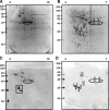
Two-dimensional gel electrophoresis and immunoblotting of elementary bodies of C. abortus. (A) A total of 75 μg of protein SYPRO Ruby stained, 3 to 10 pH range, 18-cm Immobiline DryStrip. (B) A total of 500 μg of protein SYPRO Ruby stained, 4 to 7 pH range, 18-cm Immobiline DryStrip. (C) A total of 75 μg of protein immunoblotted with serum from a selected ewe (ewe 8, 34 dpi), dilution 1:1,000, 3 to 10 pH range, 18-cm Immobiline DryStrip. Square box indicates the 26- to 32-kDa reactive antigens; the circle indicates the ∼40-kDa reactive antigens. (D) A total of 100 μg of protein immunoblotted with serum from a selected ewe (ewe 8, 34 dpi), dilution 1:1,000, 4 to 7 pH range, 18-cm Immobiline DryStrip. Circles and numbers shown in panels B and D indicate the excised protein spots identified by mass spectrometry (Table 1). Molecular mass standards (kDa) are indicated on the left.
FIG. 7.
FIG. 7.
Two-dimensional immunoblotting of elementary bodies of C. abortus. A total of 20 μg of elementary body proteins (4 to 7 pH range, 7-cm Immobiline DryStrip) were probed with sera from a pool of fetal sera (dilution 1:500). Molecular mass (kDa) standards are indicated on the left.

Similar articles

Cited by

References

    1. Azuma, Y., H. Hirakawa, A. Yamashita, Y. Cai, M. A. Rahman, H. Suzuki, S. Mitaku, H. Toh, S. Goto, T. Murakami, K. Sugi, H. Hayashi, H. Fukushi, M. Hattori, S. Kuhara, and M. Shirai. 2006. Genome sequence of the cat pathogen, Chlamydophila felis. DNA Res. 13:15-23. - PubMed
    1. Bannantine, J. P., and D. D. Rockey. 1999. Use of a primate model system to identify Chlamydia trachomatis protein antigens recognized uniquely in the context of infection. Microbiology 145:2077-2085. - PubMed
    1. Bas, S., L. Neff, M. Vuillet, U. Spenato, T. Seya, M. Matsumoto, and C. Gabay. 2008. The proinflammatory cytokine response to Chlamydia trachomatis elementary bodies in human macrophages is partly mediated by a lipoprotein, the macrophage infectivity potentiator, through TLR2/TLR1/TLR6 and CD14. J. Immunol. 180:1158-1168. - PubMed
    1. DAFF. 2006. Regional veterinary laboratories—surveillance report 2006. Department of Agriculture, Fisheries and Food, Dublin, Ireland.
    1. DAFF. 2007. Regional veterinary laboratories—surveillance report 2007. Department of Agriculture, Fisheries and Food, Dublin, Ireland.

Publication types

MeSH terms

LinkOut - more resources