. 2010 Jun 16;30(24):8083-95.
doi: 10.1523/JNEUROSCI.1091-10.2010.

A pathologic cascade leading to synaptic dysfunction in alpha-synuclein-induced neurodegeneration

Affiliations

A pathologic cascade leading to synaptic dysfunction in alpha-synuclein-induced neurodegeneration

David A Scott et al. J Neurosci. .

Abstract

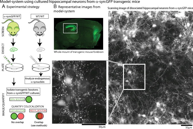
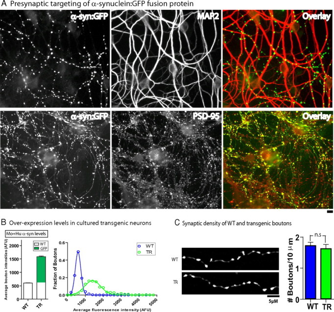
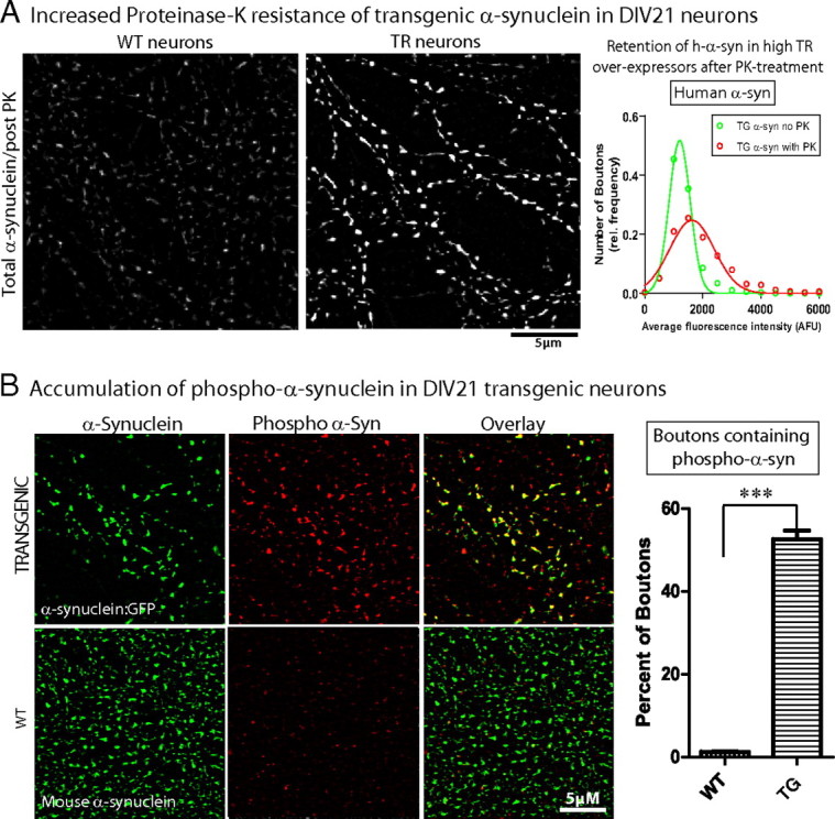
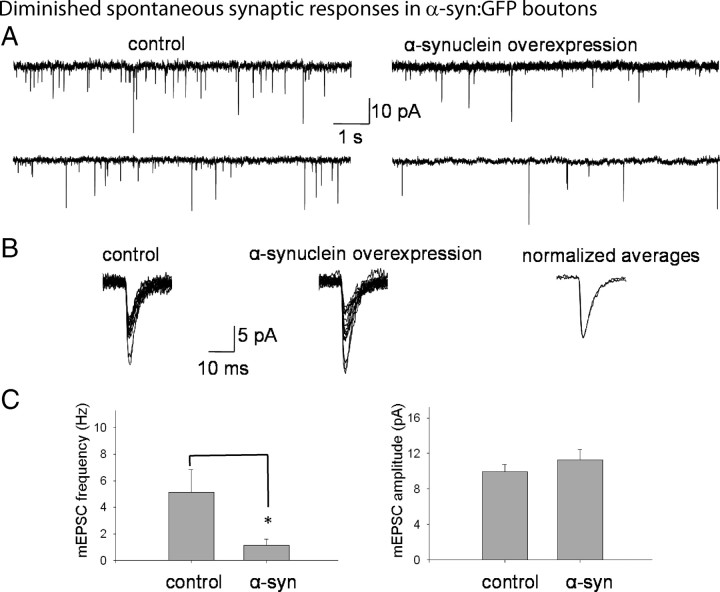
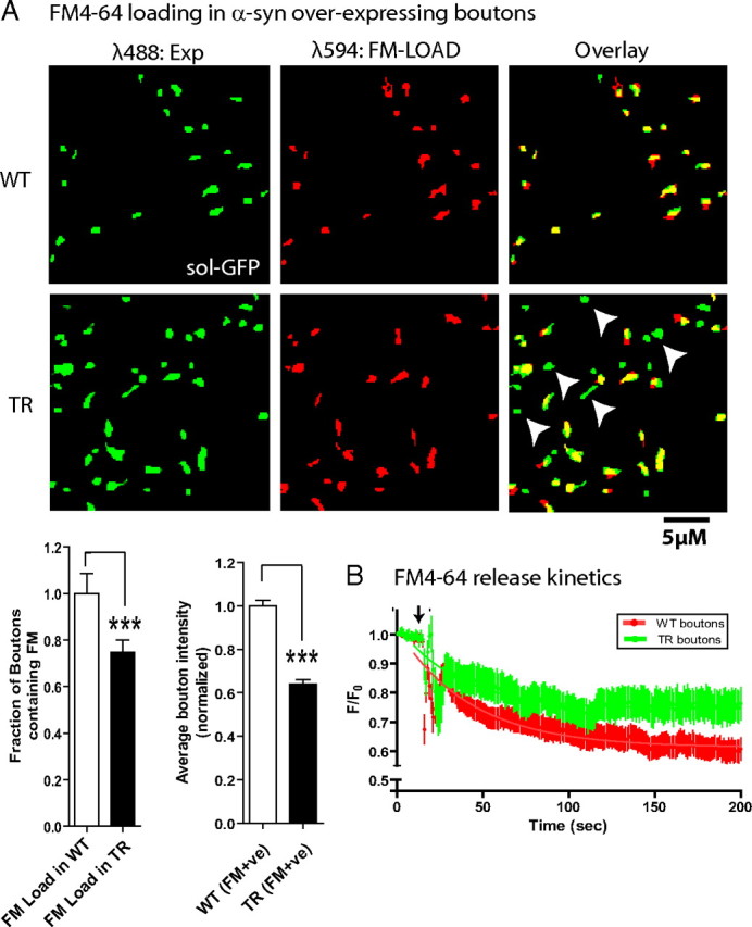
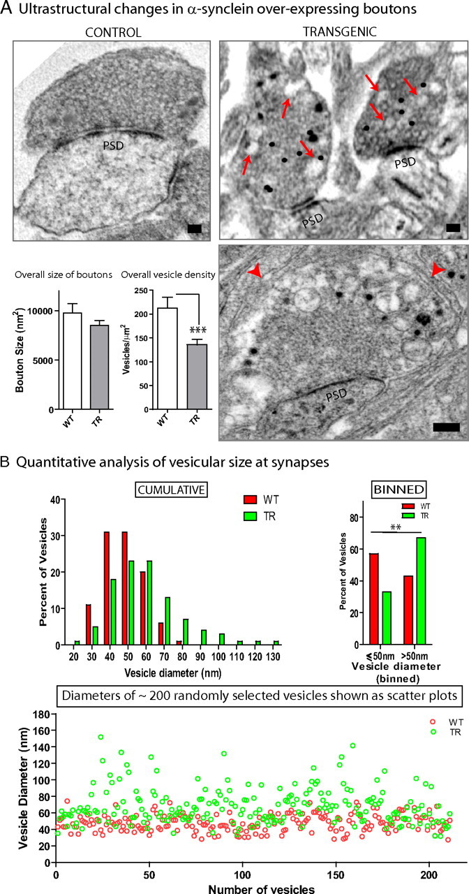
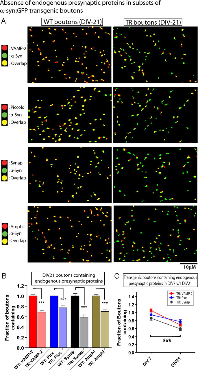
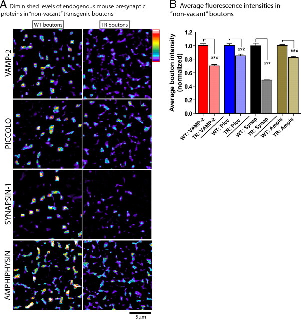
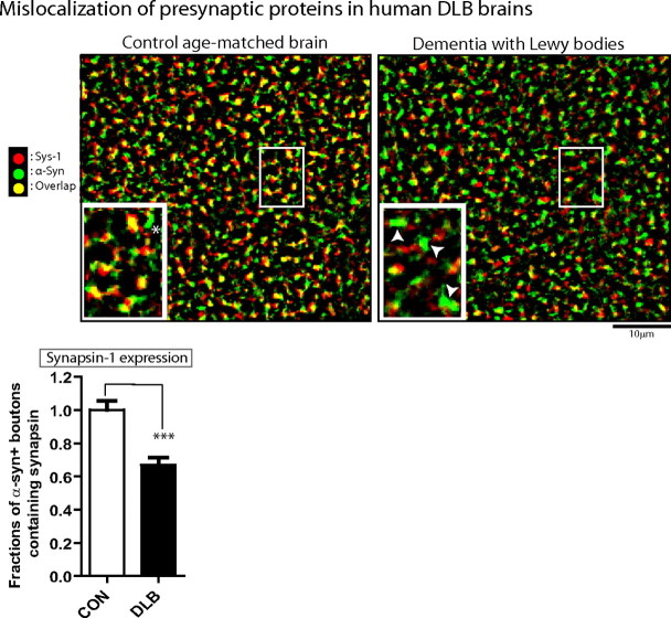
Several neurodegenerative diseases are typified by intraneuronal alpha-synuclein deposits, synaptic dysfunction, and dementia. While even modest alpha-synuclein elevations can be pathologic, the precise cascade of events induced by excessive alpha-synuclein and eventually culminating in synaptotoxicity is unclear. To elucidate this, we developed a quantitative model system to evaluate evolving alpha-synuclein-induced pathologic events with high spatial and temporal resolution, using cultured neurons from brains of transgenic mice overexpressing fluorescent-human-alpha-synuclein. Transgenic alpha-synuclein was pathologically altered over time and overexpressing neurons showed striking neurotransmitter release deficits and enlarged synaptic vesicles; a phenotype reminiscent of previous animal models lacking critical presynaptic proteins. Indeed, several endogenous presynaptic proteins involved in exocytosis and endocytosis were undetectable in a subset of transgenic boutons ("vacant synapses") with diminished levels in the remainder, suggesting that such diminutions were triggering the overall synaptic pathology. Similar synaptic protein alterations were also retrospectively seen in human pathologic brains, highlighting potential relevance to human disease. Collectively the data suggest a previously unknown cascade of events where pathologic alpha-synuclein leads to a loss of a number of critical presynaptic proteins, thereby inducing functional synaptic deficits.


Figures

Figure 1.
Figure 1.
Model system using cultured hippocampal neurons from human-α-synuclein:GFP transgenic mice. A, To develop a model system for α-synuclein overexpression, heterozygous h-α-syn:GFP+ pups (P0–P2) were optically screened (see Materials and Methods) and dissociated hippocampal neurons were plated from these mice (left); neurons from WT littermates were used as controls for all experiments (right). For quantitative studies, GFP+ (α-synuclein-overexpressing) boutons were isolated and colocalization of presynaptic proteins/dyes within the GFP+ve boutons was analyzed (see Materials and Methods for specific details of quantization algorithm). B, Top left, Native GFP fluorescence in a 3-month-old h-α-syn:GFP mouse brain section. Robust GFP expression was seen in the forebrain and hippocampus (boxed), with lower fluorescence levels throughout the neocortex. Low-power scanning representative image of native GFP fluorescence from live DIV-21 hippocampal cultures are shown. Note that numerous h-α-syn-positive punctate, bouton-like varicosities can be readily visualized in these cultures, with diffuse fluorescence in cell-bodies (magnified in inset). These methods allowed us to obtain robust α-synuclein expression in large neuronal populations and examine the evolving pathology over time.
Figure 2.
Figure 2.
Modest overexpression of human α-synuclein at synapses with no synaptic loss in DIV-21 cultured neurons. A, Transgenic h-α-syn-overexpressing neurons were fixed and stained for a somatodendritic (MAP2) and postsynaptic (PSD-95) marker. Representative images with pseudocolor overlays show apposition of h-α-syn transgenic puncta to these two markers, indicating that the vast majority of visible puncta in these cultures represent presynaptic boutons. B, Transgenic and WT DIV-21 neurons were fixed and immunostained with a pan-synuclein antibody to reveal total (mouse + human) α-synuclein. Quantitative analyses of average α-synuclein fluorescence in WT and transgenic boutons shows ∼2.5 times overexpression; distribution of data is shown as a histogram on right. C, Synaptic density was calculated by counting the number of boutons/axon length. In WT cultures, soluble GFP was transiently transfected and used as an axon/bouton-filler. No differences in presynaptic densities were seen between WT and transgenic neurons (1.7 ± 0.1 and 1.6 ± 0.1 boutons/10 μm, respectively, mean ± SEM, p = 0.63, n = 200–300 boutons analyzed for each group).
Figure 3.
Figure 3.
Pathologic alterations in DIV-21 human α-synuclein-overexpressing neurons. A, DIV-21 cultured hippocampal neurons from h-α-syn:GFP mice or their WT littermates were treated with 1 μg/ml PK as described in Materials and Methods and total (mouse + human) and human α-synuclein levels within boutons was analyzed. Focal retention of total α-synuclein (detected with GP-SYN) was seen in transgenic boutons after PK treatment as shown in the panels. Histograms of average human α-synuclein fluorescence (detected with antibody LB509) within transgenic boutons with and without PK treatment show that a larger fraction of high-α-synuclein-overexpressing boutons are present in the post-PK dataset, suggesting that boutons expressing higher h-α-syn levels are more resistant to PK treatment (average bouton intensity of human α-synuclein = 1317 ± 21.83 AFU and 1900 ± 52.20 AFU in non-PK- and PK-treated boutons, respectively, mean ± SEM; n = 700–1950 boutons). B, DIV-21 cultured hippocampal neurons from h-α-syn:GFP mice were stained with an antibody specific for the pathology-associated Ser-129 phosphorylation site of α-synuclein. While ∼53% of the transgenic boutons contained Ser-129 phosphorylated α-synuclein (top panels), negligible levels of phospho-α-synuclein was seen in WT boutons; data quantified on right (n = 500–750 boutons, ***p < 0.0001). Scale bars, lower right of images.
Figure 4.
Figure 4.
Diminished spontaneous synaptic responses in DIV-21 human α-synuclein-overexpressing neurons. DIV-21 transgenic neurons were identified by their native GFP fluorescence and electrophysiologic recordings were obtained from live neurons. For controls, WT neurons from parallel littermate cultures were analyzed. A, Representative tracings (“minis”) of spontaneous synaptic responses from hippocampal neurons overexpressing h-α-syn compared to controls. B, Individual tracings of 15 randomly selected minis from control neurons (left tracings) or h-α-syn-overexpressing neurons (middle tracings), with normalized and overlapped averages of the same events (right tracings). C, Quantitative analyses of spontaneous synaptic events. Frequencies of spontaneous events in WT and h-α-syn-overexpressing neurons were 5.20 ± 1.68 Hz and 1.15 ± 0.46 Hz, respectively, mean ± SEM, p < 0.02; n = 12–15 for each group, 500–2000 events/experiment. The amplitudes of synaptic events averaged 9.95 ± 0.8 pA and 11.47 ± 1.18 pA, and the tau decay was 6.34 ± 0.54 ms and 6.1 ± 0.68 ms in these WT and α synuclein-overexpressing neurons, respectively (mean ± SEM; *p > 0.5 for both parameters). These experiments were repeated on three separate occasions using neurons obtained from three separate litters (for each WT/transgenic group), and the data were pooled.
Figure 5.
Figure 5.
FM4-64 loading experiments. A, WT neurons transfected with soluble GFP, or h-α-syn:GFP-overexpressing neurons were loaded with 15 μm FM4-64 to label synapses, rinsed extensively, and imaged as described in Materials and Methods. First, the fraction of FM+ve boutons in the WT or transgenic group was analyzed. Left, GFP-positive boutons (green) and their corresponding FM images (red) along with the pseudocolor overlay. Note the presence of several GFP+/FM− boutons in the pseudocolor overlay from boutons overexpressing h-α-syn (some marked with arrowheads). Left graph, The fraction of transgenic boutons uptaking the FM dye relative to WT boutons was 0.748 ± 0.052 (mean ± SEM, p = 0.0115; n = 300–700 boutons), indicating that ∼25% of the boutons failed to endocytose any detectable FM dye. Right graph, Second, the average FM intensities in the boutons that did endocytose the FM dye (FM+ve) were analyzed. Average bouton intensity (in AFU) in transgenic boutons relative to WT boutons was 0.641 ± 0.021 (mean ± SEM, p < 0.0001; n = 300–500 boutons). B, Temporal kinetics of FM release. Transgenic or WT neurons were incubated with FM4-64 (see Materials and Methods), FM-loaded neurons were washed extensively, destained with high K+, and the temporal release of the dye (decay of fluorescence after stimulation) from boutons was monitored by live imaging. Decay curves from transgenic (green) and WT (red) boutons are shown; arrow marks the time point when the neurons were stimulated. F/F 0 represents the ratio of the bouton fluorescence at any given time to the initial fluorescence. The symbols and error bars are mean ± SEM, and the solid lines represent the best single-phase decay fit to the data. Note that there was a significant inhibition of FM exocytosis in the transgenic boutons, compared to boutons from their WT littermates (n = 25–40 boutons; p < 0.0001).
Figure 6.
Figure 6.
Ultrastructural alterations in DIV-21 human α-synuclein-overexpressing boutons. A, Boutons overexpressing h-α-syn:GFP were identified by immuno-EM using an antibody to GFP (immunogold signals appear as black dots) and compared to boutons from WT littermates. Top left, A typical control bouton with synaptic vesicles that are largely of uniform diameter (PSD: postsynaptic density). Top right, Two representative transgenic boutons with abnormal large vesicular profiles intermingled within synaptic vesicles of relatively normal diameters (red arrows). Bottom right, An extreme example of enlarged vesicles in transgenic synapses. Note the large vacuolar accumulations between the red arrowheads. Quantitative analyses showed that while the overall size of the boutons was unchanged, there was a significant reduction in vesicle densities within synapses (p = 0.0052; n = 5–14 boutons). Scale bar, 100 nm. B, Histograms of vesicle diameters (10 nm bins) from WT (red bars) and transgenic (green bars) highlight the variations in the overall distributions of vesicular sizes between the two vesicular populations. When the data were binned into two groups (<50 nm and >50 nm), a Fisher's exact test showed a significant increase in vesicles >50 nm in the transgenic group with a corresponding decrease in the other group (<50 nm group: 57% and 33% for WT and TR; >50 nm group: 43% and 67%, respectively; n = 200–400 vesicles). The variation in vesicular sizes within transgenic boutons is also readily visualized in the scatter plots (bottom), where vesicle diameters of ∼200 randomly selected vesicles from WT and transgenic boutons were plotted. Note that while most vesicles are ∼40–50 nm, abnormally large vesicles are far more common in the transgenic group.
Figure 7.
Figure 7.
Undetectable endogenous presynaptic proteins in subsets of h-α-syn-overexpressing boutons (“vacant synapses”). A, DIV-21 boutons overexpressing h-α-syn:GFP (green) were fixed and stained with antibodies recognizing endogenous mouse VAMP-2/piccolo/synapsin-1/amphiphysin (red). For controls, neurons from WT littermates were fixed and stained for mouse α-synuclein (green) as well as endogenous mouse VAMP-2/piccolo/synapsin-1/amphiphysin (red). The panels are pseudocolor overlays of the WT mouse (left) or the transgenic human (right) α-synucleins to the corresponding mouse endogenous proteins; colocalized boutons appear yellow. Note that while there is an almost complete colocalization of the four mouse presynaptic proteins with mouse α-synuclein in WT boutons as expected (yellow in left panels), many h-α-syn:GFP transgenic boutons did not contain any detectable presynaptic proteins (green boutons in right panels), as shown in these representative images. B, Quantification of the colocalization data. The fraction of transgenic boutons containing VAMP-2, piccolo, synapsin-1, and amphiphysin relative to WT boutons was 0.6884 ± 0.034, 0.7702 ± 0.051, 0.5902 ± 0.043, and 0.7015 ± 0.031, respectively (mean ± SEM, p < 0.0001 for all groups; n = 1000–1600 boutons/group). C, The number of boutons lacking endogenous mouse proteins increased over time, with a significantly larger numbers of boutons lacking the synaptic proteins at DIV-21, compared to DIV-7, suggesting a gradual evolution of the synaptic pathology (p < 0.0001, two-way ANOVA between groups; n = 400–700 boutons).
Figure 8.
Figure 8.
Diminished levels of presynaptic proteins in “non-vacant” transgenic boutons. Neurons were processed the same way as in Figure 7, and the analysis was focused on the transgenic boutons that did contain VAMP/Piccolo/synapsin-1/amphiphysin. A, Images on left show pseudocolor (heat maps) indicating levels of proteins within boutons, where red/white indicate higher fluorescence levels and blue/purple indicate lower (key: upper right). Note that in general, average fluorescence levels of these four proteins are higher within WT boutons, compared to transgenics. B, Average fluorescence intensities of the four mouse presynaptic proteins in transgenic boutons in transgenic boutons relative to WT boutons were 0.6992 ± 0.023, 0.8504 ± 0.026, 0.4920 ± 0.013, and 0.8250 ± 0.016, respectively (mean ± SEM, ***p < 0.001 for all groups, n = 1000–1600 boutons/each group). Scale bars, lower right of images.
Figure 9.
Figure 9.
Undetectable levels of a presynaptic protein in human brains with α-synuclein related pathology. Prefrontal cortices of autopsy human brains from controls and patients with DLB were immunostained with antibodies to h-α-syn and synapsin, and colocalization of the two antigens was evaluated. Representative images from control and LB dementia brains are shown with quantization. Note that while extensive colocalization (yellow) is evident in the control brain (left), many α-synuclein-positive boutons (green) do not contain any detectable synapsin-1 in the DLB brain (right). The area within the box was magnified (inset); arrowheads denote α-synuclein+ boutons that do not contain synapsin. The fraction of α-synuclein+ve boutons that also contained synapsin in DLB brains relative to controls was 0.6690 ± 0.046 (mean ± SEM, ***p < 0.0001; n = 2000 boutons/each group). Note that in WT brain sections, the colocalization of the various synaptic antigens was not complete (unlike cultured neurons), and there were occasional α-synuclein+ boutons in control brains that did not contain synapsin (one marked with an asterisk). Scale bars, lower right of images.

References

    1. Aarsland D, Beyer MK, Kurz MW. Dementia in Parkinson's disease. Curr Opin Neurol. 2008;21:676–682. - PubMed
    1. Abeliovich A, Schmitz Y, Fariñas I, Choi-Lundberg D, Ho WH, Castillo PE, Shinsky N, Verdugo JM, Armanini M, Ryan A, Hynes M, Phillips H, Sulzer D, Rosenthal A. Mice lacking alpha-synuclein display functional deficits in the nigrostriatal dopamine system. Neuron. 2000;25:239–252. - PubMed
    1. Anderson JP, Walker DE, Goldstein JM, de Laat R, Banducci K, Caccavello RJ, Barbour R, Huang J, Kling K, Lee M, Diep L, Keim PS, Shen X, Chataway T, Schlossmacher MG, Seubert P, Schenk D, Sinha S, Gai WP, Chilcote TJ. Phosphorylation of Ser-129 is the dominant pathological modification of alpha-synuclein in familial and sporadic Lewy body disease. J Biol Chem. 2006;281:29739–29752. - PubMed
    1. Chandra S, Fornai F, Kwon HB, Yazdani U, Atasoy D, Liu X, Hammer RE, Battaglia G, German DC, Castillo PE, Südhof TC. Double-knockout mice for alpha- and beta-synucleins: effect on synaptic functions. Proc Natl Acad Sci U S A. 2004;101:14966–14971. - PMC - PubMed
    1. Chandra S, Gallardo G, Fernández-Chacón R, Schlüter OM, Südhof TC. Alpha-synuclein cooperates with CSPalpha in preventing neurodegeneration. Cell. 2005;123:383–396. - PubMed

Publication types

MeSH terms

LinkOut - more resources