. 2010 Jun 16;30(24):8151-61.
doi: 10.1523/JNEUROSCI.0293-10.2010.

Activity-dependent bulk endocytosis and clathrin-dependent endocytosis replenish specific synaptic vesicle pools in central nerve terminals

Affiliations

Activity-dependent bulk endocytosis and clathrin-dependent endocytosis replenish specific synaptic vesicle pools in central nerve terminals

Giselle Cheung et al. J Neurosci. .

Abstract

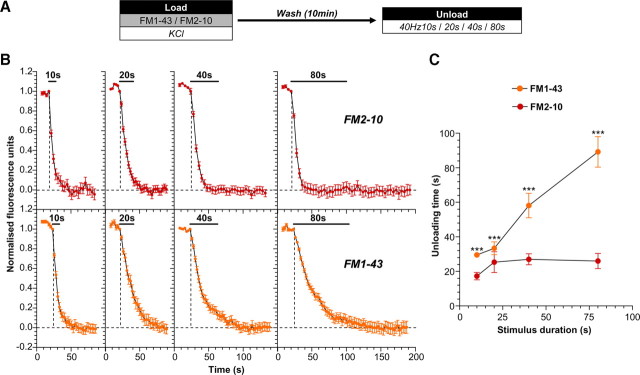
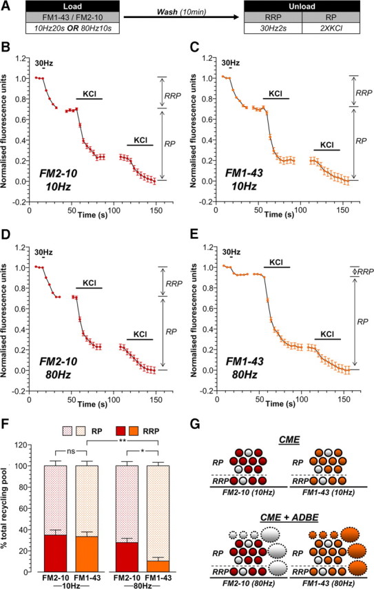
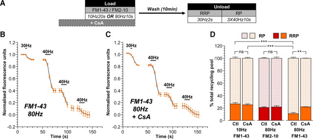
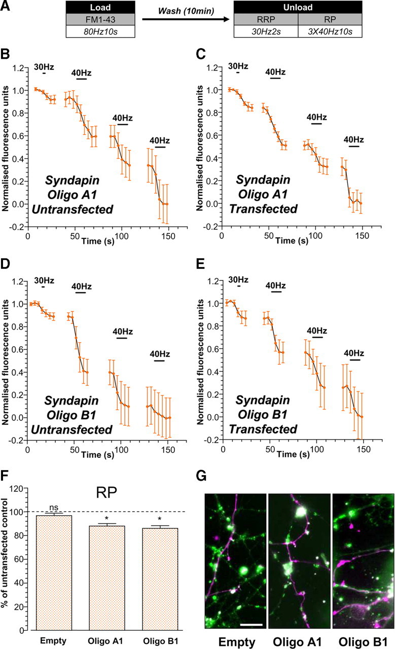
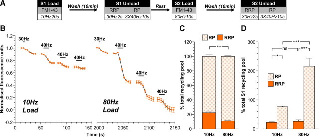
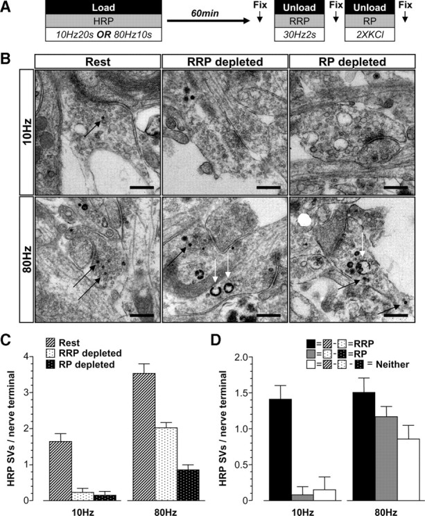
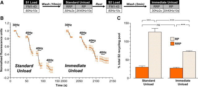
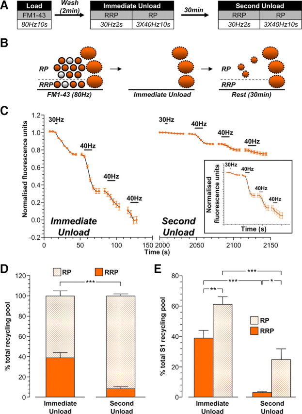
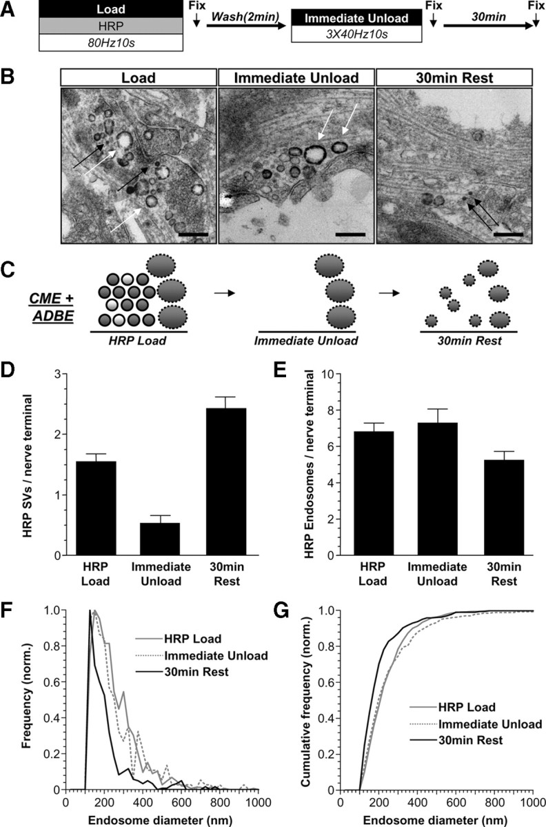
Multiple synaptic vesicle (SV) retrieval modes exist in central nerve terminals to maintain a continual supply of SVs for neurotransmission. Two such modes are clathrin-mediated endocytosis (CME), which is dominant during mild neuronal activity, and activity-dependent bulk endocytosis (ADBE), which is dominant during intense neuronal activity. However, little is known about how activation of these SV retrieval modes impact the replenishment of the total SV recycling pool and the pools that reside within it, the readily releasable pool (RRP) and reserve pool. To address this question, we examined the replenishment of all three SV pools by triggering these SV retrieval modes during both high- and low-intensity stimulation of primary rat neuronal cultures. SVs generated by CME and ADBE were differentially labeled using the dyes FM1-43 and FM2-10, and their replenishment of specific SV pools was quantified using stimulation protocols that selectively depleted each pool. Our studies indicate that while the RRP was replenished by CME-generated SVs, ADBE provided additional SVs to increase the capacity of the reserve pool. Morphological analysis of the uptake of the fluid phase marker horseradish peroxidase corroborated these findings. The differential replenishment of specific SV pools by independent SV retrieval modes illustrates how previously experienced neuronal activity impacts the capability of central nerve terminals to respond to future stimuli.


Figures

Figure 1.
Figure 1.
ADBE provides additional SVs for the total SV recycling pool. A, Cultures were loaded with either 10 μm FM1-43 or 100 μm FM2-10 in the presence of elevated KCl (50 mm) for 2 min. Dyes were washed away immediately after stimulation, and cultures were left to rest for 10 min. Dye unloading was stimulated by trains of action potentials (either 10 s, 20 s, 40 s, or 80 s) at 40 Hz. B, Representative traces of the time course of dye unloading in response to action potentials are displayed (FM2-10, red symbols, top; FM1-43, orange symbols, bottom). The duration of unloading stimulation is represented by bars. Data are normalized between 1 and 0 in all instances, where 1 = start of stimulation and 0 = cessation of unloading (indicated by dotted lines). C, The mean unloading time for either dye is plotted against the stimulus duration (FM2-10, red symbols; FM1-43, orange symbols). In all experiments, n = 4 (except 40 Hz 10 s FM1-43 and 40 Hz 80 s FM1-43, n = 3); error bars represent ±SEM; ***p < 0.001, two-way ANOVA.
Figure 2.
Figure 2.
ADBE preferentially refills the SV reserve pool. A, Cultures were loaded with either 10 μm FM1-43 or 100 μm FM2-10 using trains of either 200 action potentials (10 Hz) or 800 action potentials (80 Hz). Dyes were washed away immediately after stimulation and cultures were left to rest for 10 min. The RRP was unloaded with 60 action potentials (30 Hz), and the reserve pool (RP) was unloaded with two 30 s applications of 50 mm KCl. B–E, Representative traces of the average fluorescence drop of nerve terminals loaded with either 200 action potentials are shown (10 Hz; B, FM2-10; C, FM1-43) or 800 action potentials (80 Hz; D, FM2-10; E, FM1-43). Unloading stimulations are represented by bars. Traces were normalized (between 1 and 0) to the size of the recycling pool (RRP + RP) for each nerve terminal. The relative size of the RRP and RP are indicated by arrows. F, Average proportion of the RRP and RP as a percentage of the SV recycling pool is shown (FM2-10, red bars; FM1-43, orange bars; RRP, solid bars; RP, dotted bars). In all experiments, n = 4 (except 10 Hz FM2-10 and 80 Hz FM1-43, n = 3); error bars represent ±SEM; *p < 0.05, **p < 0.01, for both RRP and RP, one-way ANOVA. G, Schematic diagrams illustrating the extent of RRP and RP replenishment by SVs loaded with either FM2-10 (left, red) or FM1-43 (right, orange) using either 10 Hz stimulation (top, only triggers CME) or 80 Hz stimulation (bottom, triggers both CME and ADBE). Solid circles represent dye-loaded SVs; clear circles represent nonloaded SVs. Solid ovals represent dye-loaded endosomes; clear ovals represent nonloaded endosomes. Solid outlines represent CME-generated structures; dotted outlines represent ADBE-generated structures.
Figure 3.
Figure 3.
Replenishment of the reserve pool by ADBE is dependent on calcineurin activity. A, Cultures were loaded with either 10 μm FM1-43 or 100 μm FM2-10 using trains of either 200 action potentials (10 Hz) or 800 action potentials (80 Hz). Dyes were washed away immediately after stimulation and cultures were left to rest for 10 min. The RRP was unloaded with 60 action potentials (30 Hz) and the reserve pool (RP) was unloaded with three consecutive trains of 400 action potentials (40 Hz). In some experiments, cyclosporin A (CsA, 10 μm) was preincubated with cultures for 10 min before and during dye loading. B, C, Representative traces of the average fluorescence drop of nerve terminals loaded with FM1-43 evoked by 800 action potentials in the absence (Ctl) or presence of CsA are shown (B, minus CsA; C, plus CsA). Unloading stimulations are represented by bars. Traces were normalized (between 1 and 0) to the size of the recycling pool (RRP + RP) for each nerve terminal. D, Average proportion of the RRP and RP as a percentage of the SV recycling pool is shown (FM2-10, red bars; FM1-43, orange bars; RRP, solid bars; RP, dotted bars). In all experiments, n = 3 (except 10 Hz FM1-43 control, n = 4); error bars represent ±SEM; **p < 0.01, ***p < 0.001, for both RRP and RP, one-way ANOVA.
Figure 4.
Figure 4.
Replenishment of the reserve pool by ADBE is disrupted by knockdown of syndapin I expression. Cultures were transfected with either shRNA (Oligo A1, Oligo B1) against syndapin I or an empty vector 72 h before experiments. Transfected neurons were identified by mCerulean expression. A, Cultures were loaded with 10 μm FM1-43 using trains of 800 action potentials (80 Hz). Dyes were washed away immediately after stimulation and cultures were left to rest for 10 min. The RRP was unloaded with 60 action potentials (30 Hz) and the reserve pool (RP) was unloaded with three consecutive trains of 400 action potentials (40 Hz). B–E, Representative traces of the average fluorescence drop of FM1-43-loaded nerve terminals are displayed for cultures transfected with either Oligo A1 (B, C) or Oligo B1 (D, E) from both untransfected (B, D) and transfected neurons (C, E). Unloading stimuli are represented by bars. Traces were normalized (between 1 and 0) to the size of the recycling pool (RRP + RP) for each nerve terminal. F, Proportions of the RP as a percentage of the SV recycling pool were determined for each nerve terminal on transfected neurons and represented as the percentage of RP unloaded from untransfected neurons in the same experiment. In all experiments, n = 3; error bars represent ±SEM; *p < 0.05, one-sample t test with proportion of RP of untransfected neurons set as 100%. G, Fluorescent images showing FM1-43-loaded nerve terminals (FM1-43; in green) and transfected neurons (mCerulean; in magenta) for cultures transfected with empty vector (left), Oligo A1(middle), and Oligo B1 (right). Scale bar, 10 μm.
Figure 5.
Figure 5.
ADBE provides additional SVs for the reserve pool but not the RRP. A, Cultures were loaded with 10 μm FM1-43 using a train of 200 action potentials (10 Hz) at S1 and a train of 800 action potentials (80 Hz) at S2. In both cases, dyes were washed away immediately after stimulation and cultures were left to rest for 10 min. At both S1 and S2, the RRP was unloaded with a train of 60 action potentials (30 Hz) and the reserve pool (RP) was unloaded with three consecutive trains of 400 action potentials (40 Hz). Cells were left to rest for 20 min between S1 and S2. B, A representative trace of the average fluorescence drop of nerve terminals is shown (10 Hz load, S1; 80 Hz load, S2). Unloading stimulations are represented by bars. Traces were normalized (between 1 and 0) to the size of the S2 recycling pool (RRP + RP) for each nerve terminal. C, Proportion of the S1 and S2 RRP and RP as a percentage of their respective recycling pools is shown. D, Average proportion of the RRP and RP expressed as a percentage of the S1 SV recycling pool (RRP, solid bars; RP, dotted bars). In all experiments, n = 4; error bars represent ±SEM; *p < 0.05, **p < 0.01, ***p < 0.001, for both RRP and RP, one-way ANOVA.
Figure 6.
Figure 6.
Replenishment of SV pools visualized by electron microscopy. A, Cultures were loaded with 10 mg/ml HRP using either trains of 200 action potentials (10 Hz) or 800 action potentials (80 Hz). HRP was washed away immediately after stimulation and cells were left to rest for 60 min. The RRP was unloaded with 60 action potentials (30 Hz) and the reserve pool (RP) was unloaded with two 30 s applications of 50 mm KCl. Cells were fixed at three different time points indicated by arrows (rest, after RRP depletion, and after RP depletion) and processed for electron microscopy. B, Representative electron micrographs display nerve terminals that were subjected to the treatments described above. Black arrows indicate HRP-labeled SVs; white arrows indicate HRP-labeled endosomes Scale bar, 500 nm. C, HRP-labeled SVs were counted and divided by the number of nerve terminals analyzed (rest, hatched bars; RRP depleted, black dots on white bars; RP depleted, white dots on black bars). D, The size of the RRP (rest minus RRP depleted), the RP (RRP depleted minus RP depleted), and an SV population belonging to neither (rest minus both RRP depleted and RP depleted) were calculated and presented as HRP-labeled SVs per nerve terminal. Data in C and D are averages pooled from individual nerve terminals from the same experiment (10 Hz: rest n = 56 nerve terminals, RRP depleted n = 26, RP depleted n = 20; 80 Hz: rest n = 75, RRP depleted n = 112, RP depleted n = 49; all error bars represent ±SEM).
Figure 7.
Figure 7.
FM1-43-labeled SVs with delayed availability for release preferentially replenish the reserve pool. A, Cultures were loaded with 10 μm FM1-43 using a train of 800 action potentials (80 Hz) at both S1 and S2. In both cases, dyes were washed away immediately after stimulation and cultures were left to rest for either 10 min (standard unload) or 2 min (immediate unload) before unloading. At both S1 and S2, the RRP was unloaded with a train of 60 action potentials (30 Hz), and the reserve pool (RP) was unloaded with three consecutive trains of 400 action potentials (40 Hz). Cells were left to rest for 20 min between S1 and S2. B, A representative trace of the average fluorescence drop of nerve terminals is shown (standard unload, S1; immediate unload, S2). Unloading stimulations are represented by bars. Traces were normalized (between 1 and 0) to the size of the S2 recycling pool (RRP + RP) for each nerve terminal. C, Average proportion of the RRP and RP in both S1 and S2 expressed as a percentage of the S2 SV recycling pool (RRP, solid bars; RP, dotted bars). In all experiments, n = 3; error bars represent ±SEM; *p < 0.05, **p < 0.01, ***p < 0.001, one-way ANOVA.
Figure 8.
Figure 8.
SVs generated from bulk endosomes specifically replenish the reserve pool. A, Cultures were loaded with 10 μm FM1-43 using a train of 800 action potentials (80 Hz). Dye was washed away immediately after stimulation and cultures were stimulated to unload both the RRP and reserve pool (RP) after both 2 min (immediate unload) and a further 30 min (second unload). The RRP was unloaded with a train of 60 action potentials (30 Hz) and the RP was unloaded with three consecutive trains of 400 action potentials (40 Hz). B, Schematic diagram illustrating how the RRP and RP are replenished by SVs loaded with FM1-43. Solid circles represent dye-loaded SVs; clear circles represent nonloaded SVs. Solid ovals represent dye-loaded endosomes. Solid outlines represent CME-generated structures; dotted outlines represent ADBE-generated structures. C, A representative trace of the average fluorescence drop of nerve terminals is shown (immediate unload, S1; second unload, S2). Unloading stimulations are represented by bars. Traces were normalized (between 1 and 0) to the size of the S1 recycling pool (RRP + RP) for each nerve terminal. The inset displays the S2 unload normalized to the size of the S2 recycling pool. D, Proportion of the S1 and S2 RRP and RP as a percentage of their respective recycling pools is shown. E, Average proportion of the RRP and RP in both S1 and S2 expressed as a percentage of the S1 SV recycling pool (RRP, solid bars; RP, dotted bars). In all experiments n = 4; error bars represent ±SEM; *p < 0.05, **p < 0.01, ***p < 0.001, for both RRP and RP, one-way ANOVA.
Figure 9.
Figure 9.
SVs with delayed availability for release are derived from bulk and endosomes. A, Cultures were loaded with HRP (10 mg/ml) using a train of 800 action potentials (80 Hz). HRP was washed away immediately after stimulation, and after 2 min, cultures were stimulated with three consecutive trains of 400 action potentials (40 Hz) to release all available SVs. Cultures were then allowed to rest for 30 min. Cultures were fixed at three time points indicated by arrows and processed for electron microscopy. B, Representative electron micrographs display nerve terminals that were subjected to the treatments described above. Black arrows indicate HRP-labeled SVs; white arrows indicate HRP-labeled endosomes. Scale bar, 500 nm. C, Schematic diagrams illustrating nerve terminals either immediately after HRP loading (HRP load), after depletion of all available SVs (immediate unload), or after 30 min rest. Solid circles represent HRP-loaded SVs, clear circles represent nonloaded SVs. Solid ovals represent HRP-loaded endosomes. Solid outlines represent CME-generated structures, dotted outlines represent ADBE-generated structures. D, E, Mean number of HRP-labeled SVs (D) or endosomes (E) per nerve terminal is displayed for each point (data pooled from 2 independent experiments; HRP load n = 137 nerve terminals, immediate unload n = 36, 30 min rest n = 90; all error bars represent ±SEM). F, G, Endosome diameter was normalized to the total number of endosomes and plotted as frequency (F) and cumulative frequency (G) for each fixation time point (bin size = 25 nm). Only structures with average diameters ≥100 nm were considered endosomes.

References

    1. Atluri PP, Ryan TA. The kinetics of synaptic vesicle reacidification at hippocampal nerve terminals. J Neurosci. 2006;26:2313–2320. - PMC - PubMed
    1. Balaji J, Ryan TA. Single-vesicle imaging reveals that synaptic vesicle exocytosis and endocytosis are coupled by a single stochastic mode. Proc Natl Acad Sci U S A. 2007;104:20576–20581. - PMC - PubMed
    1. Clayton EL, Cousin MA. Differential labelling of bulk endocytosis in nerve terminals by FM dyes. Neurochem Int. 2008;53:51–55. - PubMed
    1. Clayton EL, Cousin MA. The molecular physiology of activity-dependent bulk endocytosis of synaptic vesicles. J Neurochem. 2009;111:901–914. - PMC - PubMed
    1. Clayton EL, Evans GJ, Cousin MA. Bulk synaptic vesicle endocytosis is rapidly triggered during strong stimulation. J Neurosci. 2008;28:6627–6632. - PMC - PubMed

Publication types

MeSH terms

LinkOut - more resources