. 2010 Jun 16;30(24):8274-84.
doi: 10.1523/JNEUROSCI.1594-10.2010.

Oxytocin-induced analgesia and scratching are mediated by the vasopressin-1A receptor in the mouse

Affiliations

Oxytocin-induced analgesia and scratching are mediated by the vasopressin-1A receptor in the mouse

Ara Schorscher-Petcu et al. J Neurosci. .

Abstract

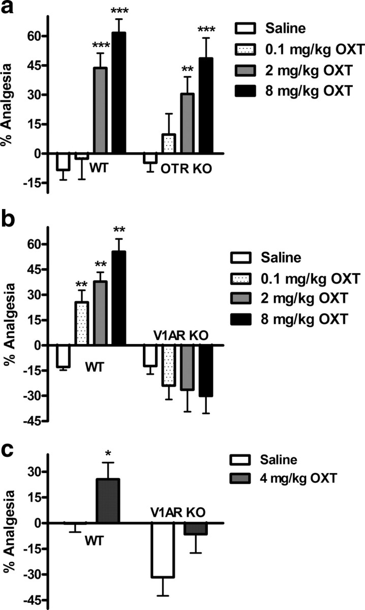
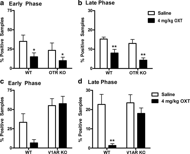
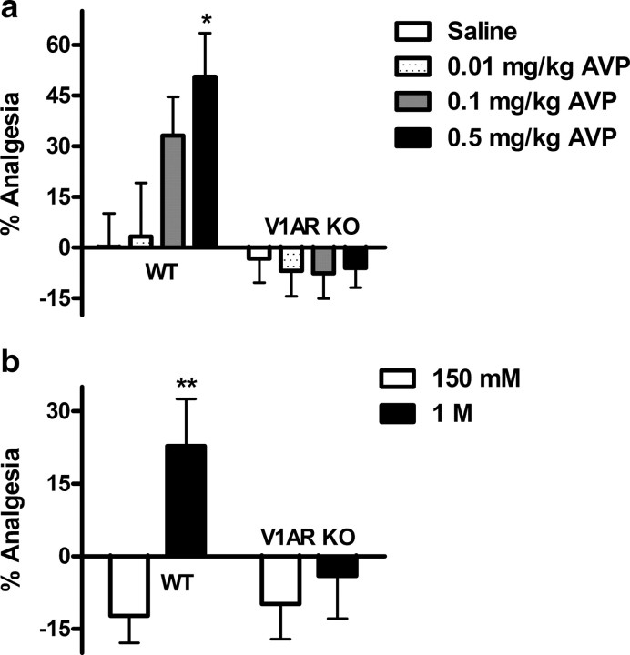
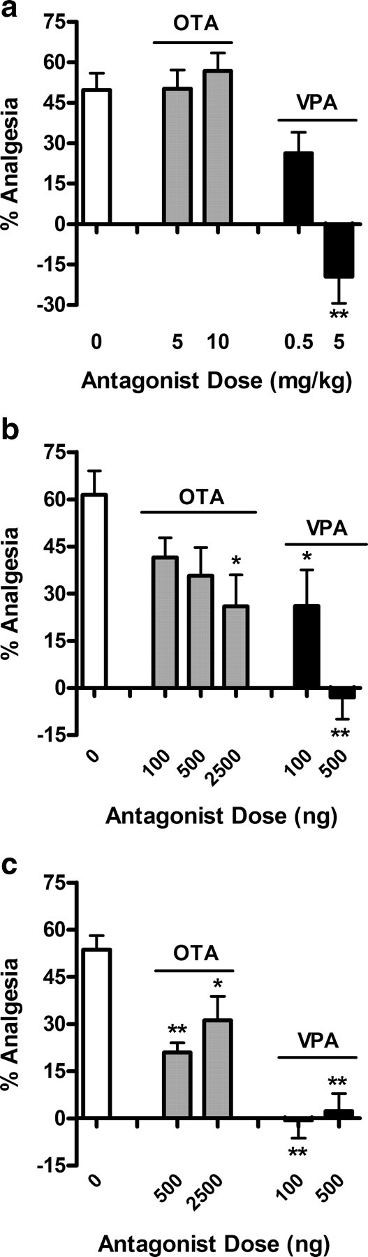
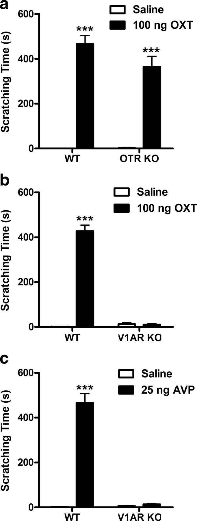
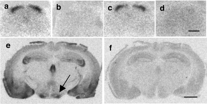
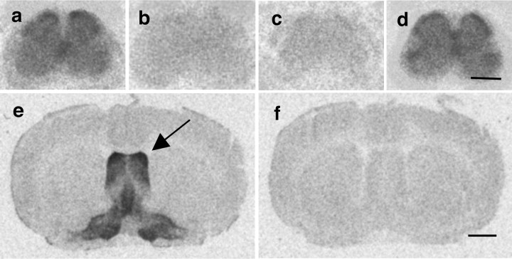
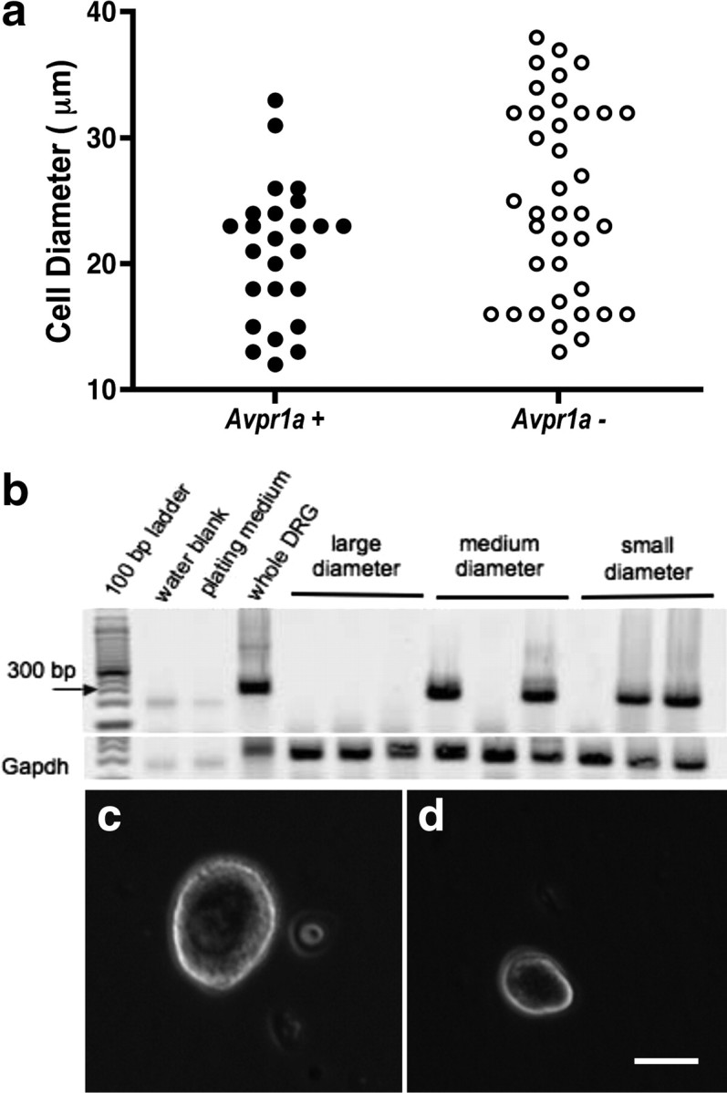
The neuropeptides oxytocin (OXT) and arginine vasopressin (AVP) contribute to the regulation of diverse cognitive and physiological functions including nociception. Indeed, OXT has been reported to be analgesic when administered directly into the brain, the spinal cord, or systemically. Here, we characterized the phenotype of oxytocin receptor (OTR) and vasopressin-1A receptor (V1AR) null mutant mice in a battery of pain assays. Surprisingly, OTR knock-out mice displayed a pain phenotype identical to their wild-type littermates. Moreover, systemic administration of OXT dose-dependently produced analgesia in both wild-type and OTR knock-out mice in three different assays, the radiant-heat paw withdrawal test, the von Frey test of mechanical sensitivity, and the formalin test of inflammatory nociception. In contrast, OXT-induced analgesia was completely absent in V1AR knock-out mice. In wild-type mice, OXT-induced analgesia could be fully prevented by pretreatment with a V1AR but not an OTR antagonist. Receptor binding studies demonstrated that the distribution of OXT and AVP binding sites in mouse lumbar spinal cord resembles the pattern observed in rat. AVP binding sites diffusely label the lumbar spinal cord, whereas OXT binding sites cluster in the substantia gelatinosa of the dorsal horn. In contrast, quantitative real-time reverse transcription (RT)-PCR revealed that V1AR but not OTR mRNA is abundantly expressed in mouse dorsal root ganglia, where it localizes to small- and medium-diameter cells as shown by single-cell RT-PCR. Hence, V1ARs expressed in dorsal root ganglia might represent a previously unrecognized target for the analgesic action of OXT and AVP.


Figures

Figure 1.
Figure 1.
OXT-induced analgesia in the radiant-heat paw withdrawal test (a, b) and von Frey test (c) of mechanical nociception is V1AR dependent. Systemically administered OXT dose-dependently produces analgesia in WT and OTR KO (a), but not in V1AR KO mice (b) (n = 7–16/group). A 4 mg/kg dose of OXT produces analgesia in WT but not V1AR KO mice (c) (n = 8/group). *p < 0.05, **p < 0.01, ***p < 0.001, compared with corresponding saline group. Error bars indicate SEM.
Figure 2.
Figure 2.
OXT-induced analgesia in the formalin test is V1AR dependent. A dose of 4 mg/kg OXT reduces licking behavior in WT and OTR KO mice in both the early (a) and late (b) phases (n = 6–8/group). In V1AR KO mice, the analgesic effect of OXT is abolished in both the early (c) and late (d) phases (n = 6–8/group). *p < 0.05, **p < 0.01, compared with corresponding saline group. Error bars indicate SEM.
Figure 3.
Figure 3.
Exogenous and endogenous AVP analgesia is V1AR dependent. AVP injected systemically is analgesic in WT, but not in V1AR KO mice (a) (n = 6–9/group). The graph in b shows the effect of osmotic stress, known to release endogenous AVP. Intraperitoneal injection of 1 m but not 150 mm (physiological) saline produces analgesia in WT but not V1AR KO mice (n = 6–10/group). *p < 0.05, compared with corresponding saline group; **p < 0.01, compared with corresponding 150 mm (physiological saline) group or other genotype. Error bars indicate SEM.
Figure 4.
Figure 4.
A selective V1AR antagonist (VPA), but not a selective OTR antagonist (OTA), prevents OXT-induced analgesia in the radiant-heat paw withdrawal test (n = 6–8/dose/drug/route). When injected systemically (a), 5 mg/kg VPA fully inhibits, whereas OTA does not affect OXT-induced analgesia. Centrally (intracerebroventricularly) administered (b), a 500 ng dose of VPA is sufficient to block OXT-induced analgesia, whereas up to 2.5 μg of OTA only partly attenuated the effect of OXT. At the spinal level (intrathecal) (c), both antagonists display some efficacy in inhibiting OXT-induced analgesia, but only the VPA is fully effective, at both 100 and 500 ng. *p < 0.05, **p < 0.01, compared with saline group. Error bars indicate SEM.
Figure 5.
Figure 5.
OXT (100 ng) administered intracerebroventricularly produces a robust and long-lasting scratching response in WT and OTR KO (a), but not in V1AR KO mice (b) (n = 7–8/group). AVP (25 ng, i.c.v.) also elicits scratching in WT, but not in V1AR KO mice (c) (n = 7–8/group). ***p < 0.001, compared with saline vehicle. Error bars indicate SEM.
Figure 6.
Figure 6.
Autoradiographic localization of specific 125I-OTA binding sites in mouse brain and spinal cord. In WT (a) and V1AR KO mice (c), OTR binding is dense in all superficial laminae of the lumbar spinal cord. OTR binding is absent in OTR KO mice (d). In WT mice, OTR binding is dense in the ventromedial hypothalamus (e, arrow). OTR binding is displaced by coincubation of radioligand with 2 μm cold OXT (b, f). Scale bars: d, 500 μm; f, 1500 μm.
Figure 7.
Figure 7.
Autoradiographic localization of specific 125I-VPA binding sites in mouse brain and spinal cord. In WT (a) and OTR KO mice (d), V1AR binding is diffuse in all regions of the lumbar spinal cord. V1AR binding is absent in V1AR KO mice (c). In WT mice, V1AR binding is dense in the lateral septum (e, arrow). V1AR binding is displaced by 2 μm cold AVP (b, f). Scale bars: d, 500 μm; f, 1500 μm.
Figure 8.
Figure 8.
Avpr1a gene expression in individual DRG cells. a, Scatterplot illustrating the proportion of Avpr1a mRNA-expressing DRG neurons of different size. b, Gel showing nine single DRG cells positive or negative for Avpr1a. c, d, Examples of large-diameter (c) and small-diameter (d) dissociated DRG neurons. Scale bar, 15 μm.

References

    1. Arnaudeau S, Leprêtre N, Mironneau J. Oxytocin mobilizes calcium from a unique heparin-sensitive and thapsigargin-sensitive store in single myometrial cells from pregnant rats. Pflugers Arch. 1994;428:51–59. - PubMed
    1. Barberis C, Tribollet E. Vasopressin and oxytocin receptors in the central nervous system. Crit Rev Neurobiol. 1996;10:119–154. - PubMed
    1. Barberis C, Balestre MN, Jard S, Tribollet E, Arsenijevic Y, Dreifuss JJ, Bankowski K, Manning M, Chan WY, Schlosser SS, Holsboer F, Elands J. Characterization of a novel, linear radioiodinated vasopressin antagonist: an excellent radioligand for vasopressin V1a receptors. Neuroendocrinology. 1995;62:135–146. - PubMed
    1. Baumgartner T, Heinrichs M, Vonlanthen A, Fischbacher U, Fehr E. Oxytocin shapes the neural circuitry of trust and trust adaptation in humans. Neuron. 2008;58:639–650. - PubMed
    1. Birnbaumer M. Vasopressin receptors. Trends Endocrinol Metab. 2000;11:406–410. - PubMed

Publication types

MeSH terms

LinkOut - more resources