. 2010 Jul;30(7):1293-9.
doi: 10.1161/ATVBAHA.108.179598.

Taking the thrombin "fork"

Affiliations

Taking the thrombin "fork"

Kenneth G Mann. Arterioscler Thromb Vasc Biol. 2010 Jul.

Abstract

The proverb that probably best exemplifies my career in research is attributable to Yogi Berra (http://www.yogiberra.com/), ie, "when you come to a fork in the road ... take it." My career is a consequence of chance interactions with great mentors and talented students and the opportunities provided by a succession of ground-breaking improvements in technology.


Figures

Fig. 1
Fig. 1. Schematic illustration of the pathways for the activation of human prothrombin
Cleavage of prothrombin at Arg273-Thr274 (Reaction 1) yields Fragment 1·2 and prethrombin 2. Further cleavage of prethrombin 2 at Arg323-Ser324 (Reaction 3) yields a disulfide-linked two-chain form of α-thrombin. Cleavage of prothrombin in the opposite order (cleavage at Arg323-Ser324; Reaction 4) yields meizothrombin. Meizothrombin is composed of the Fragment 1·2-A chain and thrombin-B chain that are covalently linked by a disulfide bond. Further cleavage at Arg273-Thr274 produces Fragment 1·2 and α-thrombin. The arrows labeled a and b indicate bonds that are susceptible to cleavage by the feedback action of thrombin. a = Arg155-Ser156; b = Arg286-Thr287. Krishnaswamy S, Church WR, Nesheim ME, Mann KG: Activation of Human Prothrombin by Human Prothrombinase. Influence of Factor Va on the Reaction Mechanism. J. Biol. Chem. 262:3291-99, 1987.
Figure 2
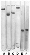
Figure 2
Electrophoretic analysis of isolated Factor V. The support medium of Gels A through D was 5% polyacrylamide. Gel A was buffered with 0.1 m Tris/borate, 5 mm CaCl2, pH 8.3. Gels B and C were buffered as described by Davis (35), and Williams and Reisfeld (36), respectively. Gel D was 0.1% in DodSO4 as described by Weber and Osborn (37). Electrophoretic analysis in DodSO4 and 20% agarose, as described by Fass et al. (38) are depicted in Gels E and F; both before E and after F reduction with 2-mercaptoethanol. Nesheim ME, Myrmel K, Hibbard L, Mann KG: Isolation and Characterization of Single Chain Bovine Factor V. J. Biol. Chem. 254:508-17, 1979.
Figure 3
Figure 3. Double reciprocal plot of the binding of Factor Xa to unstimulated platelets derived from equilibrium binding measurements of radiolabeled Factor Va and Factor Xa to platelets
The binding of Factor Xa to platelets was modeled as Factor Xa binding to platelet-bound Factor Va and can be described by Equation 1 previously detailed under “Methods.” Accordingly, the ratio of molecules of platelet-bound Factor Va to platelet-bound Factor Xa is plotted as a function of nominal Factor Xa concentration. The plot gave a dissociation constant of 6 × 10⁻¹⁰M and a binding stoichiometry of 1.04 molecules of platelet-bound Factor Va bound per molecule of bound Factor Xa. Tracy PB, Nesheim ME, Mann KG: Coordinate Binding of Factor Va and Factor Xa to the Unstimulated Platelet. J. Biol. Chem. 256:743-51, 1981.
Figure 4
Figure 4. Concepts employed in the development of the model of prothrombinase
The circle in the center represents a spherical phospholipid vesicle. This is surrounded by a region designated the interface shell (13) and is defined on the basis of the increase in hydrodynamic radius of the scattering particles that occurs upon binding of prothrombin or Factor Va to PCPS vesicles (14, 15). This region is surrounded by the bulk solution. Catalysis occurs by a mechanism that conforms to the Michaelis-Menten equation in both regions, with Km = 131 μM. In solution, the enzyme is considered Factor Xa with kcat = 0.61 min−1, whereas in the interface shell the enzyme is considered the Factor Xa-Factor Va complex with kcat = 2100 min−1 (5, 17). The rate of the reaction is given by the sum of velocities in both regions, as indicated by the equation on the figure. The last term on the right has been multiplied by δ[L]0 in order to express rates in the interface shell with respect to the total volume of the system. The term δ[L]0 is the total volume occupied in the system by the interface shell region, where [L]0 is the total phospholipid concentration and δ is a constant of proportionality. The volume occupied by the total interface shell region is typically a very small fraction (0.01%) of the total volume of the system. Although the region is “small,” it constitutes the locus in which the vast majority of prothrombin conversion occurs. The application of the model involves determination of the distribution of enzyme and substrate between bulk solution and the interface shell for any given set of initial concentrations of enzyme, substrate, and PCPS. From the distribution, bulk and interface shell concentrations are calculated, which in turn permit the calculation of expected reaction rates. Nesheim ME, Tracy RP, Mann KG: “Clotspeed,” a Mathematical Simulation of the Functional Properties of Prothrombinase. J. Biol. Chem. 259:1447-53, 1984.
Figure 5
Figure 5. Model of two collected blood coagulation reactions: conversion of factor X to factor Xa; conversion of prothrombin to thrombin. Their respective catalysts are bound adjacent to one another on their respective cofactors on the platelet membrane
Mann KG, Nesheim ME, Hibbard LS, Tracy PB: The Role of Factor V in the Assembly of the Prothrombinase Complex. In: Annals of the New York Academy of Science (Walz DA, McCoy LE, eds.), vol. 370, Contributions to Hemostasis, pp. 378-88, New York Acad. of Sci., New York, 1981.
Figure 6
Figure 6. Schematic representation of the reaction steps required for prothrombinase assembly
Prothrombinase assembly on the PCPS vesicle surface proceeds after initial reactions that yield separate Xa·PCPS and Va·PCPS binary complexes on the surface of the same vesicle. The PCPS-bound proteins then react rapidly on the vesicle surface to form prothrombinase. Krishnaswamy S, Jones KC, Mann KG: Prothrombinase Complex Assembly: Kinetic Mechanism of Enzyme Assembly on Phospholipid Vesicles. J. Biol. Chem. 263:3823-34, 1988.
Figure 7
Figure 7
Composite Western blots of fibrinogen, factor V, prothrombin, antithrombin III [AT-III], and platelet factor 4 in clotting blood. Transverse sections of Immunoblots from individual gels for various analytes separated on electrophoresis gels prepared with blood/serum samples, obtained from individual BT and subjected to tissue factor–initiated coagulation in corn trypsin inhibitor–inhibited whole blood. Gels on the same samples taken sequentially during the course of the coagulation process were run under both reducing and nonreducing conditions, transferred, and probed with the appropriate monoclonal or polyclonal antibody. Fibrinogen, factor V and factor Va light and heavy chains, and PF4 are analyzed under nonreducing conditions. Prothrombin and AT-III were analyzed under reducing conditions. At the top of the figure are indicated the time intervals at which the sample was obtained, while the bottom legend indicates the lane number on the electrophoresis gel. Clot time [4.5 minutes] is indicated by bold arrow. The letters A through H on the left margin correspond to various transverse gel sections corresponding to different analytes [see text], while the legend on the right side of the gel corresponds to the various analytes. The analytes labeled a, b, c, Tla, [TAT’], II, and d/e are not yet positively Identified. Rand MD, Lock JB, van’t Veer C, Gaffney DP and Mann KG: Blood Clotting in Minimally Altered Whole Blood. Blood 88:3432-3445, 1996.
Figure 8
Figure 8. Scheme of a two-compartment model of the regulation of TF-initiated blood coagulation
A cross-section of a blood vessel showing the luminal space, endothelial cell layer, and extravascular region is presented at the site of a perforation. The blood coagulation process in response is depicted in four stages. Tissue factor-factor VIIa complex, TF-VIIa; prothrombinase complex, Xa-Va; intrinsic factor Xase, VIIIa-IXa; ATIII-endothelial cell heparan sulfate proteoglycan complex bound to thrombin or factor Xa, HS-ATIII-(IIa or Xa); protein C bound to thrombomodulin-thrombin, TM-lia-PC. Stage 1: perforation results in delivery of blood, and with it circulating factor VIIa and platelets, to an extravascular space rich in membrane bound TF. Platelets adhere to collagen and von Willebrand factor associated with the extravascular tissue, and TF binds factor VIIa, initiating the process of factor IX and factor X activation. Factor Xa activates small amounts of prothrombin to thrombin that activates more platelets and converts factor V and factor VIII to factor Va and factor VIIIa. Stage 2: the reaction is propagated by platelet-bound intrinsic factor Xase and prothrombinase with the former being the principle factor Xa generator. Initial clotting occurs and fibrin begins to fill in the void in cooperation with activated platelets. Stage 3: a barrier composed of activated platelets ladened with procoagulant complexes and enmeshed in fibrin scaffolding is formed. The reaction in the now filled perforation is terminated by reagent consumption attenuating further thrombin generation but functional procoagulant enzyme complexes persist because they are protected from the dynamic inhibitory processes found on the intravascular face. Stage 4: view downstream of the perforation. Enzymes escaping from the plugged perforation are captured by antithrombin-heparan complexes and the protein C system is activated by residual thrombin binding to endothelial cell thrombomodulin, initiating the dynamic anticoagulant system. These intravascular processes work against occlusion of the vessel despite the continuous resupply of reactants across the intravascular face of the thrombus. Orfeo T, Butenas S, Brummel-Ziedins KE, Mann KG. J Biol Chem. The tissue factor requirement in blood coagulation. J. Biol. Chem. 280:42887-42896, 2005.

Similar articles

Cited by

References

    1. Fish WW, Mann KG, Tanford C. The estimation of polypeptide chain molecular weights by gel filtration in 6 M guanidine hydrochloride. J Biol Chem. 1969;244:4989–4994. - PubMed
    1. Mann KG, Batt CW. The molecular weights of bovine thrombin and its primary autolysis products. J Biol Chem. 1969;244:6555–6557. - PubMed
    1. Mann KG, Yip R, Heldebrant CM, Fass DN. Multiple active forms of thrombin. 3. Polypeptide chain location of active site serine and carbohydrate. J Biol Chem. 1973;248:1868–1875. - PubMed
    1. Weber K, Osborn M. The reliability of molecular weight determinations by dodecyl sulfate-polyacrylamide gel electrophoresis. J Biol Chem. 1969;244:4406–4412. - PubMed
    1. Davie EW, Ratnoff OD. Waterfall sequence for intrinstic blood clotting. Science. 1964;145:1310–1312. - PubMed

Publication types

Personal name as subject