. 2010 Jul 15;185(2):1248-57.
doi: 10.4049/jimmunol.1000289. Epub 2010 Jun 16.

Viral infection of the placenta leads to fetal inflammation and sensitization to bacterial products predisposing to preterm labor

Affiliations

Viral infection of the placenta leads to fetal inflammation and sensitization to bacterial products predisposing to preterm labor

Ingrid Cardenas et al. J Immunol. .

Erratum in

  • J Immunol. 2011 Sep 1;187(5):2835. Booth, Carmen [corrected to Booth, Carmen J]

Abstract

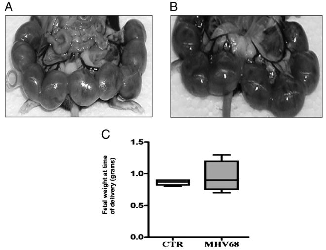
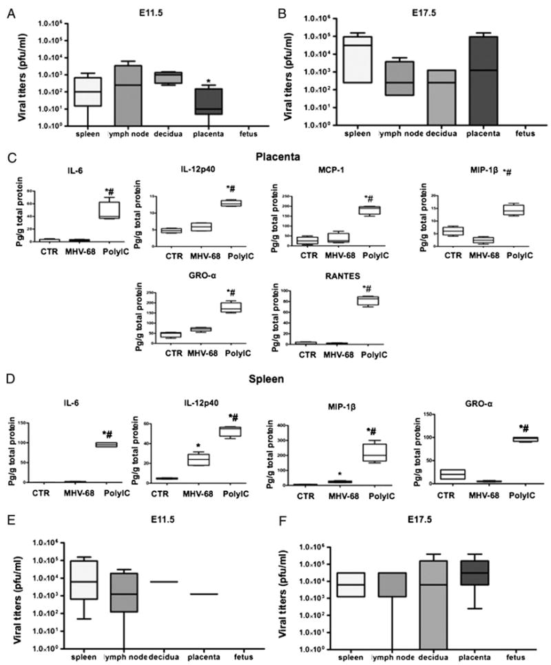
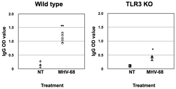
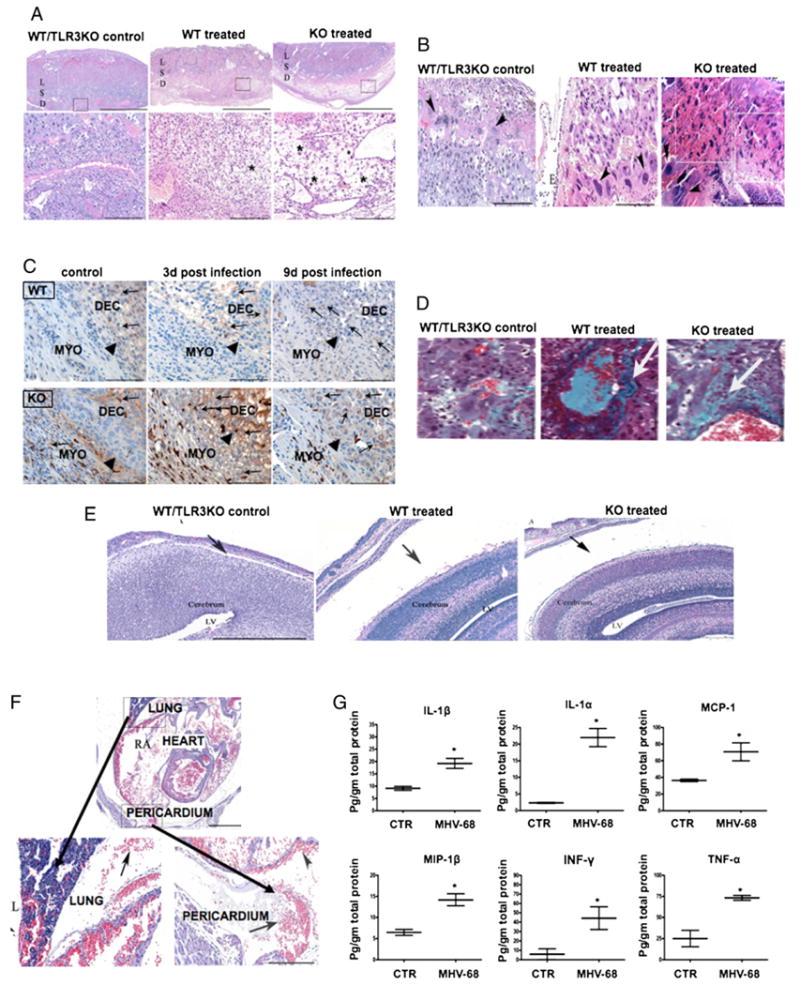
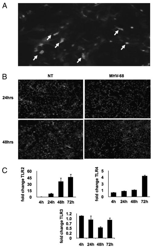
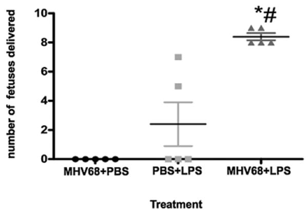
Pandemics pose a more significant threat to pregnant women than to the nonpregnant population and may have a detrimental effect on the well being of the fetus. We have developed an animal model to evaluate the consequences of a viral infection characterized by lack of fetal transmission. The experiments described in this work show that viral infection of the placenta can elicit a fetal inflammatory response that, in turn, can cause organ damage and potentially downstream developmental deficiencies. Furthermore, we demonstrate that viral infection of the placenta may sensitize the pregnant mother to bacterial products and promote preterm labor. It is critical to take into consideration the fact that during pregnancy it is not only the maternal immune system responding, but also the fetal/placental unit. Our results further support the immunological role of the placenta and the fetus affecting the global response of the mother to microbial infections. This is relevant for making decisions associated with treatment and prevention during pandemics.


Conflict of interest statement

Disclosures: The authors have no financial conflicts of interest.

Figures

FIGURE 1
FIGURE 1
Pregnancy outcome in wt mice infected with MHV-68. Wt pregnant mice were infected i.p. with 1 × 10⁶ PFU MHV-68 or vehicle at E8.5 and sacrificed at E17.5. A, Pups, uterus, and gestational sacs from wt treated with vehicle, and B, pups, uterus, and gestational sac from wt infected with MHV-68, showing no differences in gross anatomy. C, Fetal weight at the time of delivery. Note the lack of difference between the two groups. n = 6 mice per group.
FIGURE 2
FIGURE 2
Effect of MHV-68 viral infection in pregnant mice. Viral titers as PFU/ml were determined in wt pregnant mice infected with MHV-68 (1 × 10⁶ PFU) 3 d (E11.5; A) and 9 d (E17.5; B) postinfection. Viral titers were observed in lymph nodes, placenta, decidua, and spleen, but were absent in the fetuses. *p < 0.05, decidua versus spleen. Placenta (C) and spleen (D) cytokine profile was determined in wt pregnant mice treated with poly(I:C) or MHV-68 4 and 9 d postinfection, respectively. *p < 0.05, MHV-68 versus control; #p < 0.05, poly(I:C) versus MHV-68. E and F, Viral titers as PFU/ml were determined in TLR-3 KO pregnant mice infected with MHV-68: E, 3 d (E11.5), and F, 9 d (E17.5) postinfection. Note the high levels of viral titers in lymph nodes, placenta, decidua, and spleen, but absent in the fetuses. Bars show median ± SEM. n = 6 mice per group.
FIGURE 3
FIGURE 3
Seroconversion in wt and TLR-3 KO pregnant mice infected with MHV-68. Wt and TLR-3 KO mice were infected i.p. with MHV-68 (1 × 10⁶ PFU) or vehicle at E8.5. Serum samples were collected 9 dpi, and levels of IgG Abs were determined by ELISA. Note the high levels of IgG anti–MHV-68 Abs in the wt treated group compared with controls. A significantly lower response was observed in TLR-3 KO-treated mice. n = 6 mice per group. *p < 0.05.
FIGURE 4
FIGURE 4
Effect of MHV-68 viral infection on the maternal/fetal interface. Morphological changes were observed in the placenta and decidua of MHV-68–infected pregnant mice associated with the following: A, edema (*) in the D, but absent in the L and S. Upper and lower panel, scale bar, 200 and 300 μm, respectively. B, Necrosis in placenta, marked loss of cellular detail, fragmentation, hypereosinophilia (boxes) in the labyrinth subjacent to the epithelium (E), and necrosis of scattered giant cells (arrowheads). These changes were more accentuated in the TLR-3 KO compared with wt mice. Scale bars, 200 μm. C, Immunohistochemistry for F4/80-positive macrophages (brown) localized in the myometrium (MYO) and decidua (DEC) at E11.8 and E17.5. Black arrows show the edge between myometrium and decidua. Original magnification ×20. D, Increase in collagen deposition (arrows) in the labyrinth layer of MHV-68–infected mice using Trichromic Mason staining (original magnification ×20. E, Presence of fetal brain hydrocephalus (black arrows) in wt and TLR-3 KO MHV-68–infected mice (middle and right panel) compared with normal controls (left panel). Note the width of LVs. Original magnification ×10. F, Fetal thoracic cavity from WT and TLR-3 KO pregnant mice infected with MHV-68. Note the areas of hemorrhage in lung right middle lobe and pericardium. G, Fetal cytokine profile from pregnant mice infected with MHV-68. Fetal lysates were obtained, and cytokines/chemokines were measured by Luminex. Bars show median ± SEM. n = 6 mice per group. *p < 0.05. Figures are representative of six animals per group and three independent experiments. D, decidua; L, labyrinth; LV, lateral ventricle; S, sponginous layer.
FIGURE 5
FIGURE 5
Primary cultures of human first trimester trophoblast cells infected with GFP-MHV-68. A, Infection was monitored by the presence of GFP-labeled MHV-68. Positive GFP-MHV-68–infected trophoblast cells (white arrows) were observed ≈12 h postinfection. B, Inhibition of NF-κB activity in MHV-68–infected trophoblast cells. Expression of p65 was determined by immunofluorescence. Note the decrease in the number of trophoblast cells with nuclear p65 (white dots) following MHV-68 infection. C, Expression of TLR-2, -3, and -4 by human first trimester trophoblast cells following MHV-68 infection. TLR-2, -3, and -4 expressions were determined by real-time quantitative RT-PCR. Note the significant increase on TLR-2 and -4 expression and decrease in TLR-3 in MHV-68–infected cells compared with the control. n = 3 samples per group. *p < 0.05.
FIGURE 6
FIGURE 6
MHV-68 infection sensitizes to bacterial LPS. Wt mice were infected i.p. with MHV-68 at E8.5, followed by a single dose of LPS (20 μg/kg) at E15.5. LPS induced preterm labor in all of the animals that received prior MHV-68 infections (triangles), compared with animals receiving only LPS (squares) or MHV-68 infection (circles). Bars show median ± SEM. n = 6 mice per group. *p < 0.05. MHV-68 plus LPS versus PBS plus LPS. #p < 0.05. MHV-68 plus LPS versus PBS plus MHV-68.

References

    1. Nuzum JW, Pilot I, Stangl FH, Bonar BE. 1918 pandemic influenza and pneumonia in a large civil hospital. IMJ Ill Med J. 1976;150:612–616. - PubMed
    1. Romero R, Espinoza J, Chaiworapongsa T, Kalache K. Infection and prematurity and the role of preventive strategies. Semin Neonatol. 2002;7:259–274. - PubMed
    1. Jamieson DJ, Honein MA, Rasmussen SA, Williams JL, Swerdlow DL, Biggerstaff MS, Lindstrom S, Louie JK, Christ CM, Bohm SR, et al. Novel Influenza A (H1N1) Pregnancy Working Group H1N1 2009 influenza virus infection during pregnancy in the USA. Lancet. 2009;374:451–458. - PubMed
    1. Burguete T, Rabreau M, Fontanges-Darriet M, Roset E, Hager HD, Köppel A, Bischof P, Schlehofer JR. Evidence for infection of the human embryo with adeno-associated virus in pregnancy. Hum Reprod. 1999;14:2396–2401. - PubMed
    1. Basurko C, Carles G, Youssef M, Guindi WE. Maternal and fetal consequences of dengue fever during pregnancy. Eur J Obstet Gynecol Reprod Biol. 2009;147:29–32. - PubMed

Publication types

MeSH terms