. 2010 Aug;151(8):4024-30.
doi: 10.1210/en.2009-1251. Epub 2010 Jun 16.

Multiple endocrine neoplasia type 1 deletion in pancreatic alpha-cells leads to development of insulinomas in mice

Affiliations

Multiple endocrine neoplasia type 1 deletion in pancreatic alpha-cells leads to development of insulinomas in mice

H-C Jennifer Shen et al. Endocrinology. 2010 Aug.

Abstract

The pancreatic alpha- and beta-cells are critical components in regulating blood glucose homeostasis via secretion of glucagon and insulin, respectively. Both cell types are typically localized in the islets of Langerhans. However, little is known about the roles of paracrine interactions that contribute to their physiological functions. The lack of suitable cell lines to study alpha- and beta-cells interactions have led us to develop an alpha-cell-specific Cre-expressing transgenic line utilizing a glucagon promoter sequence, the Glu-Cre transgenic mouse. Here, we demonstrate that the Glu-Cre could specifically and efficiently excise floxed target genes in adult islet alpha-cells. We further showed that deletion of the tumor suppressor gene, multiple endocrine neoplasia type 1 (Men1), in alpha-cells led to tumorigenesis. However, to our surprise, the lack of Men1 in alpha-cells did not result in glucagonomas but rather beta-cell insulinomas. Because deletion of the Men1 alleles was only present in alpha-cells, our data suggested that cross communication between alpha- and beta-cells contributes to tumorigenesis in the absence of Men1. Together, we believed that the new model systems described here will allow future studies to decipher cellular interactions between islet alpha- and beta-cells in a physiological context.


Figures

Figure 1
Figure 1
Histological analyses of Cre recombinase expression in pancreatic islets. A, Immunofluorescent staining of Cre recombinase using a Cre antibody in Glu-Cre;Men1 f/+ mice at 6 wk of age. Left, Insulin staining (green) was used to identify islet, and glucagon (red) was used to confirm presence of α-cells. Right, Closely adjacent frozen section was used to identify Cre-positive cells (red) in the same islet, 4′,6-diamidino-2-phenylindole (DAPI) (blue) nuclear stain was used to visualize the islet morphology. B, Using the Glu-Cre;Z/AP reporter mice at 5 months of age, AP and glucagon (dark brown) staining of islets are shown, with insets indicated zoom-in positive staining for each.
Figure 2
Figure 2
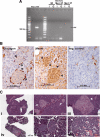
Generation of Glu-Cre;Men1 f/f mice. A, Deletion of the Men1 alleles in 4-month-old mouse pancreas. Genotyping PCR using genomic DNA isolated from pancreatic endocrine insulin-positive β-cells (I) and glucagon-positive α-cells (G). The presence of deleted Men1 allele (del; 638 bp) was only detected in Glu-Cre;Men1 f/f, but not in wild type (Wt) and Men1 f/f pancreas. Positive (pos) and negative (neg) controls are as indicated. B, Glucagon and menin protein expression in Glu-Cre;Men1 f/f islets at 13 months of age by immunohistochemical staining. Dark brown color indicated positive cytoplasmic staining for glucagon (left) and nuclear staining for menin (middle, noting nonspecific light brown staining in both endocrine and exocrine tissues). Negative control (neg. control) is shown to demonstrate background staining without glucagon and menin antibodies. Arrowheads pointed to examples of glucagon-positive cells lacking menin, in which cell nuclei lack the dark brown positive menin staining in the middle panel. C, Hematoxylin and eosin staining of representative Glu-Cre;Men1 f/f animals with hyperplastic islets (i) and tumors (iv) at 13–14 months of age. Normal-sized islets (ii and v) are indicated by arrows, whereas hyperplastic islets (iii) and insulinomas tumors (vi) filled with blood islands are indicated by arrowheads.
Figure 3
Figure 3
Representative immunohistological staining of insulin (A–D) and glucagon (E–H) on Glu-Cre;Men1 f/f mouse pancreas. The entire pancreas section stained for insulin (A) and glucagon (E) is shown. Scale bars, 0.5 mm. B–D and F–H, Magnified areas of A and E. Scale bars, 0.05 mm, except for D and H, where scale bars, 0.1 mm. For the glucagon staining, nonspecific light brown background color was observed in some islets.
Figure 4
Figure 4
Analysis of 14- to 16-month-old Glu-Cre;Men1 f/f pancreatic islets. A, Deletion of the Men1 alleles in islet glucagon-positive α-cells (G), but not in insulin-positive β-cells (I), in two Glu-Cre;Men1 f/f mice. Control genotypes (Glu-Cre and Men1 f/+), positive (pos) and negative (neg) controls are as indicated. B, Slight increase in the percentage of glucagon cells in pancreatic islets of Glu-Cre;Men1 f/f mice (n = 4), when compare with age-matched Glu-Cre mice (n = 2).

Similar articles

Cited by

References

    1. Gromada J, Franklin I, Wollheim CB 2007 α-Cells of the endocrine pancreas: 35 years of research but the enigma remains. Endocr Rev 28:84–116 - PubMed
    1. Cabrera O, Berman DM, Kenyon NS, Ricordi C, Berggren PO, Caicedo A 2006 The unique cytoarchitecture of human pancreatic islets has implications for islet cell function. Proc Natl Acad Sci USA 103:2334–2339 - PMC - PubMed
    1. Bonner-Weir S, Orci L 1982 New perspectives on the microvasculature of the islets of Langerhans in the rat. Diabetes 31:883–889 - PubMed
    1. Chandrasekharappa SC, Guru SC, Manickam P, Olufemi SE, Collins FS, Emmert-Buck MR, Debelenko LV, Zhuang Z, Lubensky IA, Liotta LA, Crabtree JS, Wang Y, Roe BA, Weisemann J, Boguski MS, Agarwal SK, Kester MB, Kim YS, Heppner C, Dong Q, Spiegel AM, Burns AL, Marx SJ 1997 Positional cloning of the gene for multiple endocrine neoplasia-type 1. Science 276:404–407 - PubMed
    1. Lemmens I, Van de Ven WJ, Kas K, Zhang CX, Giraud S, Wautot V, Buisson N, De Witte K, Salandre J, Lenoir G, Pugeat M, Calender A, Parente F, Quincey D, Gaudray P, De Wit MJ, Lips CJ, Höppener JW, Khodaei S, Grant AL, Weber G, Kytölä S, Teh BT, Farnebo F, Thakker RV 1997 Identification of the multiple endocrine neoplasia type 1 (MEN1) gene. The European consortium on MEN1. Hum Mol Genet 6:1177–1183 - PubMed

Publication types