

Severe In vivo hyper-homocysteinemia is not associatedwith elevation of amyloid-beta peptides in the Tg2576 mice
- PMID: 20555139
- PMCID: PMC3880572
- DOI: 10.3233/JAD-2010-100171
Severe In vivo hyper-homocysteinemia is not associatedwith elevation of amyloid-beta peptides in the Tg2576 mice
Abstract
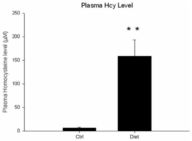
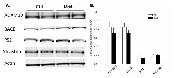
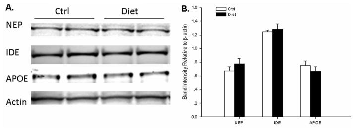
Since hyper-homocysteinemia (HHcy) was recognized as a risk factor for Alzheimer's disease (AD), many studies tried to induce HHcy in animal models to investigate its effect on amyloid-beta protein precursor (AbetaPP) metabolism. Previous reports found that HHcy induced in AD transgenic mouse models, by either feedina a methionine-enriched diet or vitamin Bs deficient diet, is associated with elevation of amyloid-beta (Abeta) levels. However, there is no data available on the effect of dietary intervention which combines both excessive methionine and low levels of vitamin Bs on amyloidogenesis in any of these models. In the current study, we investigated the effect of a combination diet, which was both enriched in methionine and deficient in folate, vitamin B6 and B12, in an AD mouse model, the Tg2576. We found that 7 months treatment of this diet induced severe HHcy in these mice with plasma homocysteine level higher than 150 microM. However, no difference was detected in brain Abeta levels or deposition between the diet-treated and control group. As shown by western blot, severe HHcy did not alter the steady state levels of proteins involved in AbetaPP metabolism, either. These results demonstrate that this combination diet-induced severe HHcy does not influence amyloidogenesis in vivo.
Figures


Similar articles
-
Acceleration of brain amyloidosis in an Alzheimer's disease mouse model by a folate, vitamin B6 and B12-deficient diet.Exp Gerontol. 2010 Mar;45(3):195-201. doi: 10.1016/j.exger.2009.12.005. Epub 2009 Dec 11. Exp Gerontol. 2010. PMID: 20005283 Free PMC article.
-
Folate/Vitamin B Alleviates Hyperhomocysteinemia-Induced Alzheimer-Like Pathologies in Rat Retina.Neurosci Bull. 2019 Apr;35(2):325-335. doi: 10.1007/s12264-018-0293-8. Epub 2018 Sep 28. Neurosci Bull. 2019. PMID: 30264378 Free PMC article.
-
Diet-induced hyperhomocysteinemia increases amyloid-beta formation and deposition in a mouse model of Alzheimer's disease.Curr Alzheimer Res. 2010 Mar;7(2):140-9. doi: 10.2174/156720510790691326. Curr Alzheimer Res. 2010. PMID: 19939226 Free PMC article.
-
Transthyretin accelerates vascular Abeta deposition in a mouse model of Alzheimer's disease.Brain Pathol. 2009 Jan;19(1):48-57. doi: 10.1111/j.1750-3639.2008.00166.x. Epub 2008 Apr 22. Brain Pathol. 2009. PMID: 18429966 Free PMC article.
-
Is hyperhomocysteinemia an Alzheimer's disease (AD) risk factor, an AD marker, or neither?Trends Pharmacol Sci. 2011 Sep;32(9):562-71. doi: 10.1016/j.tips.2011.05.003. Epub 2011 Jun 20. Trends Pharmacol Sci. 2011. PMID: 21684021 Free PMC article. Review.
Cited by
-
Protective Effects of Dendrobium nobile Lindl. Alkaloids on Alzheimer's Disease-like Symptoms Induced by High-methionine Diet.Curr Neuropharmacol. 2022;20(5):983-997. doi: 10.2174/1570159X19666210809101945. Curr Neuropharmacol. 2022. PMID: 34370639 Free PMC article.
-
Impact of Hyperhomocysteinemia and Different Dietary Interventions on Cognitive Performance in a Knock-in Mouse Model for Alzheimer's Disease.Nutrients. 2020 Oct 23;12(11):3248. doi: 10.3390/nu12113248. Nutrients. 2020. PMID: 33114054 Free PMC article.
-
Characterization of human S-adenosyl-homocysteine hydrolase in vitro and identification of its potential inhibitors.J Enzyme Inhib Med Chem. 2017 Dec;32(1):1209-1215. doi: 10.1080/14756366.2017.1370584. J Enzyme Inhib Med Chem. 2017. PMID: 28933241 Free PMC article.
-
Mechanistic Link between Vitamin B12 and Alzheimer's Disease.Biomolecules. 2022 Jan 14;12(1):129. doi: 10.3390/biom12010129. Biomolecules. 2022. PMID: 35053277 Free PMC article. Review.
-
β-amyloid deposition is shifted to the vasculature and memory impairment is exacerbated when hyperhomocysteinemia is induced in APP/PS1 transgenic mice.Alzheimers Res Ther. 2014 Jun 9;6(3):32. doi: 10.1186/alzrt262. eCollection 2014. Alzheimers Res Ther. 2014. PMID: 24991237 Free PMC article.
References
-
- Huang T, Yuan G, Zhang Z, Zou Z, Li D. Cardiovascular pathogenesis in hyperhomocysteinemia. Asia Pac J Clin Nutr. 2008;17:8–16. - PubMed
-
- Boushey CJ, Beresford SA, Omenn GS, Motulsky AG. A quantitative assessment of plasma homocysteine as a risk factor for vascular disease. Probable benefits of increasing folic acid intakes. JAMA. 1995;274:1049–1057. - PubMed
-
- Seshadri S, Beiser A, Selhub J, Jacques PF, Rosenberg IH, D’Agostino RB, Wilson PW, Wolf PA. Plasma homocysteine as a risk factor for dementia and Alzheimer’s disease. N Engl J Med. 2002;346:476–483. - PubMed
-
- Clarke R, Smith AD, Jobst KA, Refsum H, Sutton L, Ueland PM. Folate, vitamin B12, and serum total homocysteine levels in confirmed Alzheimer disease. Arch Neurol. 1998;55:1449–1455. - PubMed
-
- Seshadri S. Elevated plasma homocysteine levels: risk factor or risk marker for the development of dementia and Alzheimer’s disease? J Alzheimers Dis. 2006;9:393–398. - PubMed
Publication types
MeSH terms
Substances
Grants and funding
LinkOut - more resources
Full Text Sources
Medical
Research Materials
