

Brain perfusion correlates of visuoperceptual deficits in mild cognitive impairment and mild Alzheimer's disease
- PMID: 20555146
- PMCID: PMC3306804
- DOI: 10.3233/JAD-2010-091069
Brain perfusion correlates of visuoperceptual deficits in mild cognitive impairment and mild Alzheimer's disease
Abstract
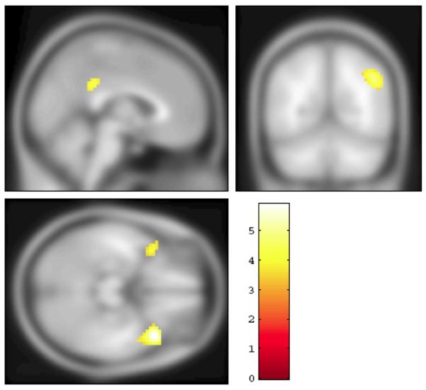
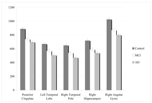
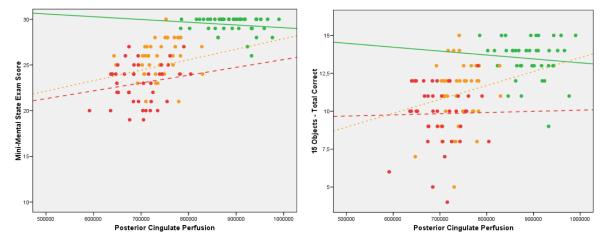
Visuoperceptual processing is impaired early in the clinical course of Alzheimer's disease (AD). The 15-Objects Test (15-OT) detects such subtle performance deficits in mild cognitive impairment (MCI) and mild AD. Reduced brain perfusion in the temporal, parietal, and prefrontal regions have been found in early AD and MCI patients. The objectives of this study were to confirm the role of the 15-OT in the diagnosis of MCI and AD and to investigate the brain perfusion correlates of visuoperceptual dysfunction (15-OT) in subjects with MCI, AD, and normal aging. Forty-two AD, 42 MCI, and 42 healthy elderly control subjects underwent a brain Single Photon Emission Tomography (SPECT) and separately completed the 15-OT. An analysis of variance compared 15-OT scores between groups. SPM5 was used to analyse the SPECT data. 15-OT performance was impaired in the MCI and AD patients. In terms of the SPECT scans, AD patients showed reduced perfusion in temporal-parietal regions, while the MCI subjects had decreased perfusion in the middle and posterior cingulate. When MCI and AD groups were compared, a significant brain perfusion reduction was found in temporo-parietal regions. In the whole sample, 15-OT performance was significantly correlated with the clinical dementia rating scores, and with the perfusion in the bilateral posterior cingulate and the right temporal pole, with no significant correlation in each separate group. Our findings suggest that the 15-OT performance provides a useful gradation of impairment from normal aging to AD, and it seems to be related to perfusion in the bilateral posterior cingulate and the right temporal pole.
Figures
References
-
- Bell-McGinty S, Lopez OL, Meltzer CC, Scanlon JM, Whyte EM, DeKosky ST, Becker JT. Differential cortical atrophy in subgroups of mild cognitive impairment. Arc Neurol. 2005;62:1393–1397. - PubMed
-
- Devanand DP, Pradhaban G, Liu X, Khandji A, De Santi S, Segal S, Rusinek H, Pelton GH, Honig LS, Mayeux R, Stern Y, Tabert MH, de Leon MJ. Hippocampal and entorhinal atrophy in mild cognitive impairment: prediction of Alzheimer disease. Neurology. 2007;68:828–836. - PubMed
-
- Whitwell JL, Josephs KA, Murray ME, Kantarci K, Przybelski SA, Weigand SD, Vemuri P, Senjem ML, Parisi JE, Knopman DS, Boeve BF, Petersen RC, Dickson DW, Jack CR. MRI correlates of neurofibrillary tangle pathology at autopsy: A voxel-based morphometry study. Neurology. 2008;71:743–749. - PMC - PubMed
Publication types
MeSH terms
Grants and funding
LinkOut - more resources
Full Text Sources
Medical
