. 2010 Jul 27;49(29):6206-18.
doi: 10.1021/bi100728q.

Structural basis for hydration dynamics in radical stabilization of bilin reductase mutants

Affiliations

Structural basis for hydration dynamics in radical stabilization of bilin reductase mutants

Amanda C Kohler et al. Biochemistry. .

Abstract

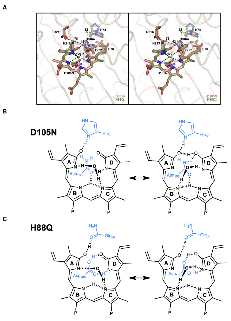
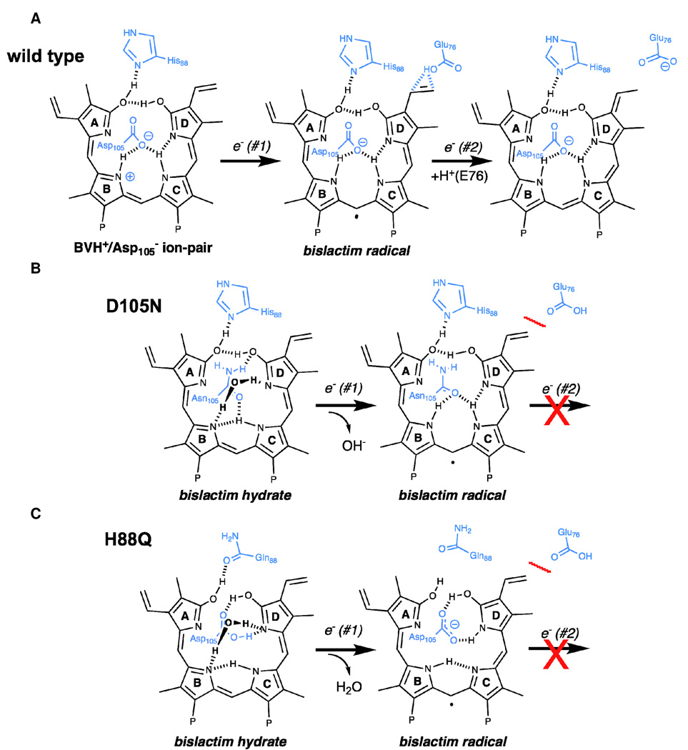
Heme-derived linear tetrapyrroles (phytobilins) in phycobiliproteins and phytochromes perform critical light-harvesting and light-sensing roles in oxygenic photosynthetic organisms. A key enzyme in their biogenesis, phycocyanobilin:ferredoxin oxidoreductase (PcyA), catalyzes the overall four-electron reduction of biliverdin IXalpha to phycocyanobilin--the common chromophore precursor for both classes of biliproteins. This interconversion occurs via semireduced bilin radical intermediates that are profoundly stabilized by selected mutations of two critical catalytic residues, Asp105 and His88. To understand the structural basis for this stabilization and to gain insight into the overall catalytic mechanism, we report the high-resolution crystal structures of substrate-loaded Asp105Asn and His88Gln mutants of Synechocystis sp. PCC 6803 PcyA in the initial oxidized and one-electron reduced radical states. Unlike wild-type PcyA, both mutants possess a bilin-interacting axial water molecule that is ejected from the active site upon formation of the enzyme-bound neutral radical complex. Structural studies of both mutants also show that the side chain of Glu76 is unfavorably located for D-ring vinyl reduction. On the basis of these structures and companion (15)N-(1)H long-range HMQC NMR analyses to assess the protonation state of histidine residues, we propose a new mechanistic scheme for PcyA-mediated reduction of both vinyl groups of biliverdin wherein an axial water molecule, which prematurely binds and ejects from both mutants upon one electron reduction, is required for catalytic turnover of the semireduced state.


Figures

Figure 1
Figure 1
PcyA Reaction. (A) PcyA catalyzes the four-electron reduction of biliverdin (BV) to phycocyanobilin (PCB) via the intermediacy of 181,182-dihydrobiliverdin (15). Four proton-coupled-electron-transfers are mediated by stepwise one-electron transfers from four reduced ferredoxins (Fd). Pyrrole rings are labeled A – D, ‘P’ represents the propionate groups, and the small numbers indicate carbon numbering for BV. (B) Different tautomeric forms of Asp105 with the D-ring pyrrole for wild-type PcyA. The major ‘bi-dentate’ neutral conformation observed as 65% occupancy (19) is depicted both neutral lactam and lactim tautomers. The minor ‘axial’ ion-pair conformation is shown on the right. Dashed lines represent hydrogen bonds. The positive charge of the minor conformation on the B-ring also can be delocalized on other pyrrole rings and on His88.
Figure 2
Figure 2
Structural models for BV-bound D105N and H88Q mutants of Synechocystis PcyA. (A) Overall structures of both mutants in their ‘oxidized’ states are indistinguishable from those of wild-type PcyA. The protein backbone is depicted as a rainbow-shaded ribbon cartoon colored blue at the N-terminus and red at the C-terminus. Bound BV is shown in a ball-and-stick representation with white-colored carbon atoms. Cyan-colored electron density drawn at 1.0 sigma level for BV in the active site of ‘oxidized’ (B) H88Q and (C) D105N mutants. Orange-colored electron density is contoured at 3 sigma from an annealed omit map (only waters omitted) revealing three tightly bound water molecules, one of which (i.e. Wat400) is missing in wild-type PcyA (19). (D) Wat400 is lost upon reduction of D105N (as shown) and H88Q (not shown) mutants.
Figure 3
Figure 3
Active sites of PcyA D105N and H88Q mutants. (A) D105N (salmon) and H88Q (green) mutants superimpose with an RMSD of 0.172Å for 242 equivalent alpha-carbons. BV substrate, conserved water molecules, and side chains of key residues are modeled in the active site. Many residues display multiple conformations (see text). Dashed lines denote potential hydrogen bonds in the D105N structure (3.5Å H-bond distance cutoff) and ordered solvent molecules are colored in the same color as the carbons. (B) Mono-dentate lactim and lactam complexes of the BV D-ring bound to the D105N mutant. (C) Bi-dentate lactim and lactam complexes of the BV D-ring bound to the H88Q mutant. Protein residues are drawn in blue.
Figure 4
Figure 4
Structural comparison of semi-reduced bilin radical complexes of PcyA D105N and H88Q mutants with their oxidized states and with each other. (A) D105N oxidized (salmon color) and radical (sand color) structures. (B) H88Q oxidized (green) to H88Q radical (cyan) structures. (C) H88Q and D105N radical structures (coloring same as above). Water 400 departs in both radical structures.
Figure 5
Figure 5
2D 15N-1H LR-HMQC spectra of BV-free (A) and BV-bound (B) PcyA at pH 7.0. Spectra of wild type (black), H88Q (green) and D105N (red) are overlaid. Peaks at 8.55 and 6.30 ppm in (A) are assigned to His88. The peak at 7.12 ppm (marked by an asterisk) is assigned to His74 (data not shown). Dashed lines in red connect the set of resonances assigned to H88 in the BV-bound D105N mutant. Complete chemical shift assignments are given in Supplemental Table 1 (see text for discussion).
Figure 6
Figure 6
Proposed structural models for one electron reduction of wild-type, D105N and H88Q PcyA. (A) The BVH+/Asp ion pair accepts the initial electron in wild-type PcyA to generate a neutral radical intermediate. Glu76 is ideally positioned for the second proton-couple electron transfer. For both D105N (B) and H88Q (C) mutants, BV is not protonated and Glu76 is poorly positioned for secondary electron transfer. For D105N, one electron transfer to enzyme-bound substrate is accompanied by proton transfer from Wat400 and ejection of hydroxide ion. For H88Q, proton-coupled electron transfer mediated by Asp105 instead results in ejection of water. Protein residues are drawn in blue.
Figure 7
Figure 7
A revised catalytic mechanism for wild-type PcyA is based on structural data. See Discussion for details.

Similar articles

Cited by

References

    1. McDonagh AF. Bile Pigments: Bilatrienes and 5,15-Biladienes. In: Dolphin D, editor. The Porphyrins. New York: Academic Press; 1979. pp. 293–491.
    1. Ortiz de Montellano PR, Auclair K. Heme Oxygenase Structure and Mechanism. In: Guilard R, editor. The Porphyrin Handbook. The Iron and Cobalt Pigments: Biosynthesis, Structure and Degradation. New York City: Academic Press; 2003. pp. 183–210.
    1. Beale SI. Biosynthesis of phycobilins. Chemical Reviews. 2002;93:785–802.
    1. Frankenberg N, Lagarias JC. Biosynthesis and Biological Function of Bilins. In: Guilard R, editor. The Porphyrin Handbook. Chlorophylls and Bilins: Biosynthesis Structure and Degradation. New York: Academic Press; 2003. pp. 211–235.
    1. Rockwell NC, Su YS, Lagarias JC. Phytochrome structure and signaling mechanisms. Ann. Rev. Plant Biol. 2006;57:837–858. - PMC - PubMed

Publication types