Comparative Study
. 2010 Sep 1;114(5):1487-97.
doi: 10.1111/j.1471-4159.2010.06864.x. Epub 2010 Jun 16.

Altered neurotransmission in the mesolimbic reward system of Girk mice

Affiliations
Comparative Study

Altered neurotransmission in the mesolimbic reward system of Girk mice

Devinder Arora et al. J Neurochem. .

Abstract

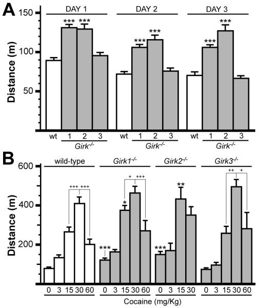
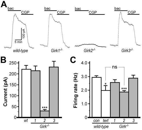
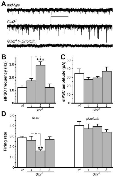
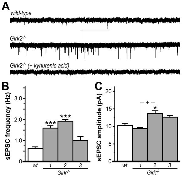
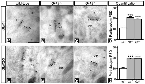
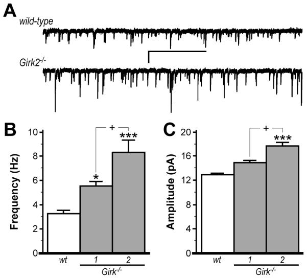
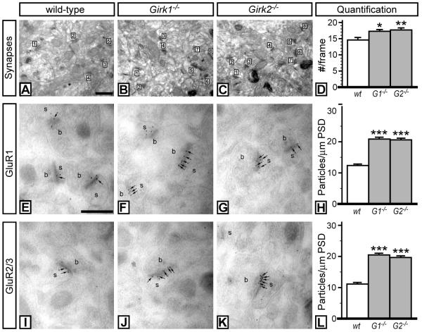
Mice lacking the Girk2 subunit of G protein-gated inwardly rectifying K+ (Girk) channels exhibit dopamine-dependent hyperactivity and elevated responses to drugs that stimulate dopamine neurotransmission. The dopamine-dependent phenotypes seen in Girk2(-/-) mice could reflect increased intrinsic excitability of or diminished inhibitory feedback to midbrain dopamine neurons, or secondary adaptations triggered by Girk2 ablation. We addressed these possibilities by evaluating Girk(-/-) mice in behavioral, electrophysiological, and cell biological assays centered on the mesolimbic dopamine system. Despite differences in the contribution of Girk1 and Girk2 subunits to Girk signaling in midbrain dopamine neurons, Girk1(-/-) and Girk2(-/-) mice exhibited comparable baseline hyperactivities and enhanced responses to cocaine. Girk ablation also correlated with altered afferent input to dopamine neurons in the ventral tegmental area. Dopamine neurons from Girk1(-/-) and Girk2(-/-) mice exhibited elevated glutamatergic neurotransmission, paralleled by increased synaptic levels of alpha-amino-3-hydroxyl-5-methyl-4-isoxazole-propionate glutamate receptors. In addition, synapse density, alpha-amino-3-hydroxyl-5-methyl-4-isoxazole-propionate receptor levels, and glutamatergic neurotransmission were elevated in medium spiny neurons of the nucleus accumbens from Girk1(-/-) and Girk2(-/-) mice. We conclude that dopamine-dependent phenotypes in Girk2(-/-) mice are not solely attributable to a loss of Girk signaling in dopamine neurons, and likely involve secondary adaptations facilitating glutamatergic signaling in the mesolimbic reward system.


Figures

Figure 1
Figure 1. Baseline and cocaine-induced motor activity in wild-type and Girk−/− mice
A) Total distance traveled (m) for wild-type (wt), Girk1−/−, Girk2−/−, and Girk3−/− mice in open-field environments during 1-h sessions on 3 consecutive days (n=36-81 per genotype). Statistical symbols: *** − P<0.001, vs. wild-type (within day). B) Cocaine-induced motor activity in wild-type and Girk−/− mice. Subjects were challenged with saline or one of 4 cocaine doses (3, 15, 30, 60 mg/kg i.p.) and total distance traveled (m) was measured during the 60-min post-injection window. Cocaine triggered dose-dependent increases in motor activity in wild-type (F4,125=54.6; P<0.001), Girk1−/− (F4,130=65.3; P<0.001), Girk2−/− (F3,52=14.4; P<0.001), and Girk3−/− (F4,81=65.3; P<0.001) mice. Genotype-dependent differences in cocaine-induced responses were observed following injection of 0 (F3,192=23.0; P<0.001) and 15 mg/kg cocaine (F3,87=6.1; P<0.001), but not after the 3 (F3,41=2.0; P=0.1), 30 (F3,43=1.8; P=0.2), or 60 (F3,41=2.0; P=0.1) mg/kg injections (n=6-30 per genotype and dose). Statistical symbols: *, **, *** − P<0.05, 0.01, and 0.001, respectively, vs. wild-type (within dose); within-genotype comparisons are displayed to convey differences observed at the higher cocaine doses: +, ++, +++ − P<0.05, 0.01, and 0.001, respectively, relative to the response at 30 mg/kg cocaine.
Figure 2
Figure 2. Functional characterization of VTA DA neurons from wild-type and Girk−/− mice
A) Outward currents evoked by baclofen (bac, 200 μM) in VTA DA neurons from wild-type, Girk1−/−, Girk2−/−, and Girk3−/− mice. Outward currents corresponded with a decrease in input resistance and were sensitive to 0.3 external Ba2+ (not shown), and were reversed by the GABABR antagonist CGP54626 (CGP, 2 μM). B) Summary of baclofen-induced outward currents in VTA DA neurons from wild-type (219±17 pA), Girk1−/− (212±21 pA), Girk2−/− mice (31±4 pA), and Girk3−/− (229±37 pA) mice (n=6-22 per genotype). Genotype-dependent differences in peak baclofen-induced current amplitude were evident (F3,39=11.9, P<0.001). Symbols: ***, P<0.001 vs. wild-type. C) A significant effect of group on firing rate was observed (F4,147=10.5, P<0.001) with respect to ex vivo firing frequencies of VTA DA neurons from wild-type mice at baseline (con, 2.93±0.12 Hz) and following application of the Girk channel blocker tertiapin (tert, 1.98±0.34 Hz), and at baseline in slices from Girk1−/− (2.55±0.20 Hz), Girk2−/− mice (1.84±0.11 Hz) and Girk3−/− (2.87±0.22 Hz) mice (n=9-49 per group). Note that there was no significant (ns) difference in firing rates of VTA DA neurons from Girk2−/− mice and wild-type neurons treated with tertiapin. Symbols: *, ***, P<0.05 and 0.001, respectively, vs. wild-type.
Figure 3
Figure 3. Inhibitory input to VTA DA neurons in wild-type and Girk−/− mice
A) Representative traces of sIPSCs measured in VTA DA neurons from wild-type (upper) and Girk2−/− mice (middle); sIPSCs were blocked by the GABA receptor antagonist picrotoxin (100 μM, lower). Scale bars: 40 pA/5s. Summary histograms of sIPSC frequency and amplitude in VTA DA neurons from wild-type (wt, 1.2±0.2 Hz, 34.4±5.7 pA), Girk1−/− (1.7±0.2 Hz, 26.9±3.1 pA), Girk2−/− (2.9±0.3 Hz, 29.2±2.2 pA), and Girk3−/− (1.2±0.3 Hz, 36.6±5.5 pA), mice are shown in panels B and C, respectively (n=7-10 per genotype). A significant effect of genotype was observed for sIPSC frequency (F3,29=10.1: P<0.001) but not amplitude (F3,29=1.1: P=0.4). Symbols: *** P<0.001 vs. wild-type and Girk3−/−; + P<0.05, Girk1−/− vs. Girk2−/−. D) Firing frequency of VTA DA neurons from wild-type and Girk−/− mice (n=8-21 per genotype) were measured at baseline (left) and following administration of 100 μM picrotoxin (right). An effect of genotype on firing rates was observed at baseline (F3,56=10.0; P<0.001) but not after picrotoxin application (F3,56=1.0: P=0.4). Symbols: ** P<0.01 vs. wild-type and Girk3−/−; + P<0.05, Girk1−/− vs. Girk2−/−.
Figure 4
Figure 4. Excitatory input to VTA DA neurons in wild-type and Girk−/− mice
A) Representative traces of sEPSCs measured in VTA DA neurons from wild-type (upper) and Girk2−/− mice (middle); sEPSCs were blocked by the non-selective ionotropic glutamate receptor antagonist kynurenic acid (2 mM; lower). Scale bars: 20 pA/5 s. Summary histograms of sEPSC frequency and amplitude in VTA DA neurons from wild-type (wt, 0.6±0.1 Hz, 10.4±0.8 pA), Girk1−/− (1.6±0.1 Hz, 8.9±0.4 pA), Girk2−/− (1.9±0.1 Hz, 13.5±0.8 pA), and Girk3−/− (1.0±0.2 Hz, 12.7±0.4 pA), mice are shown in panels B and C, respectively (n=7-12 per genotype). A significant effect of genotype was observed for both sEPSC frequency (F3,31=18.4; P<0.001) and amplitude (F3,31=8.8; P<0.001). Symbols: *,*** P<0.05 and 0.001, respectively, vs. wild-type; + P<0.001, Girk1−/− vs. Girk2−/−.
Figure 5
Figure 5. AMPA subunit labeling in VTA DA neurons from wild-type and Girk−/− mice
Electron micrographs showing GluR1 (A-C) and GluR2/3 (E-G) immunoreactivities in VTA DA neurons from wild-type, Girk1−/−, and Girk2−/− mice, as detected using double-labeling, post-embedding immunogold electron microscopy. In sections from wild-type mice, immunoparticles for GluR1 (A) and GluR2/3 (E) subunits (arrows, 10 nm gold particles) were found along the PSDs of dendritic shafts (Den) of DA neurons (TH-positive cells, arrowheads, 20 nm gold particles) establishing contact with axon terminals (b). In sections from Girk1−/− mice (B,F) and Girk2−/− mice (C,G), an increase in the number of immunoparticles for both GluR1 and GluR2/3 subunits along the PSD was observed. These images are representative of data from three separate panels of wild-type and Girk−/− mice. Scale bar: 0.2 μm. D,H) Quantification of GluR1 and GluR2/3 synaptic density in VTA DA neurons from wild-type, Girk1−/− (G1−/−), and Girk2−/− (G2−/−) mice. Synaptic density is expressed as number of immunoparticles per 1 μm length of PSD. Symbols: *** P<0.001 vs. wild-type.
Figure 6
Figure 6. Excitatory input to NAcc medium spiny neurons in wild-type and Girk−/− mice
A) Representative traces of mEPSCs in NAcc DA neurons from wild-type (upper) and Girk2−/− (lower) mice. Scale bars: 2 s/5 pA. mEPSCs were blocked completely by the non-selective ionotropic glutamate receptor antagonist kynurenic acid (2 mM, not shown). Summary of mEPSC frequency and amplitude for medium spiny neurons in the NAcc shell from wild-type (wt, 3.3±0.3 Hz, 12.9±0.4 pA), Girk1−/− (5.5±0.4 Hz, 14.6±0.7 pA), and Girk2−/− (8.3±1.0 Hz, 17.1±0.8 pA) mice are shown in panels B and C, respectively (n=11-23 per genotype). Genotype-dependent differences were observed for both mEPSC frequency (F2,44=21.8; P<0.001) and amplitude (F2,44=14.6; P<0.001). Symbols: *,*** P<0.05 and 0.001, respectively, vs. wild-type; + P<0.05, Girk1−/− vs. Girk2−/−.
Figure 7
Figure 7. Markers of excitatory neurotransmission in the NAcc of wild-type and Girk−/− mice
Electron micrographs taken from the NAcc core of wild-type (A), Girk1−/− (B), and Girk2−/− (C) mice, showing excitatory synapses (numbered) in the neuropil. Synapses between dendritic spines and axon terminals that exhibited a clear PSD were counted. Scale bar: 0.2 μm. D) Quantification of synapses from wild-type, Girk1−/− (G1−/−), and Girk2−/− (G2−/−) mice. The number of synapses per frame (#/frame) is plotted as a function of genotype. Synapses were tabulated in 21 frames per genotype, from 3 different animals per genotype. A significant effect of genotype on synapse density was observed (F2,60=5.4; P<0.01). Symbols: *,** P<0.05 and 0.01, respectively, vs. wild-type. E-L) AMPA subunit labeling in NAcc medium spiny neurons from wild-type and Girk−/− mice. Electron micrographs showing GluR1 (E-G) and GluR2/3 (I-K) immunoreactivity in medium spiny neurons from wild-type, Girk1−/−, and Girk2−/− mice, as detected using post-embedding immunogold electron microscopy. In sections from wild-type mice, immunoparticles for GluR1 (E) and GluR2/3 (I) subunits (arrows, 10 nm gold particles) were found along PSDs on individual spines (s) in contact with axon terminals (b). In sections from Girk1−/− (F,J) and Girk2−/− (G,K) mice, an increase in the number of immunoparticles for both GluR1 and GluR2/3 subunits along the PSD was observed. Scale bar: 0.2 μm. H,L) Quantification of GluR1 and GluR2/3 synaptic density in Nacc medium spiny neurons from wild-type, Girk1−/− (G1−/−), and Girk2−/− (G2−/−) mice. Synaptic density is expressed as number of immunoparticles per 1 μm length of PSD. Symbols: *** P<0.001 vs. wild-type.

References

    1. Beckstead MJ, Grandy DK, Wickman K, Williams JT. Vesicular dopamine release elicits an inhibitory postsynaptic current in midbrain dopamine neurons. Neuron. 2004;42:939–946. - PubMed
    1. Bettahi I, Marker CL, Roman MI, Wickman K. Contribution of the Kir3.1 subunit to the muscarinic-gated atrial potassium channel IKACh. J Biol Chem. 2002;277:48282–48288. - PubMed
    1. Bjorklund A, Dunnett SB. Dopamine neuron systems in the brain: an update. Trends Neurosci. 2007;30:194–202. - PubMed
    1. Blednov YA, Stoffel M, Chang SR, Harris RA. GIRK2 deficient mice. Evidence for hyperactivity and reduced anxiety. Physiol Behav. 2001;74:109–117. - PubMed
    1. Blednov YA, Stoffel M, Cooper R, Wallace D, Mane N, Harris RA. Hyperactivity and dopamine D1 receptor activation in mice lacking girk2 channels. Psychopharmacology (Berl) 2002;159:370–378. - PubMed

Publication types

MeSH terms

Substances