

Patterns of the Cranial Venous System from the Comparative Anatomy inVertebrates. Part III. The Ventricular System and Comparative Anatomy of the Venous Outlet of Spinal Cord and Its Homology with the Five Brain Vesicles
- PMID: 20557753
- PMCID: PMC3313715
- DOI: 10.1177/159101990801400203
Patterns of the Cranial Venous System from the Comparative Anatomy inVertebrates. Part III. The Ventricular System and Comparative Anatomy of the Venous Outlet of Spinal Cord and Its Homology with the Five Brain Vesicles
Abstract
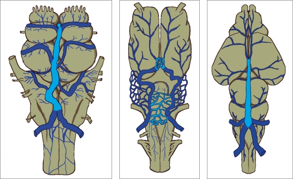
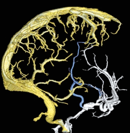
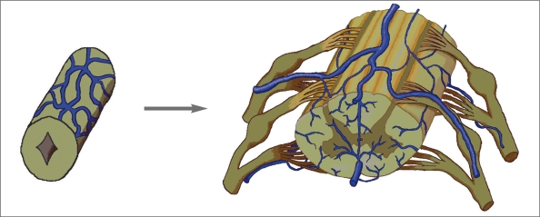
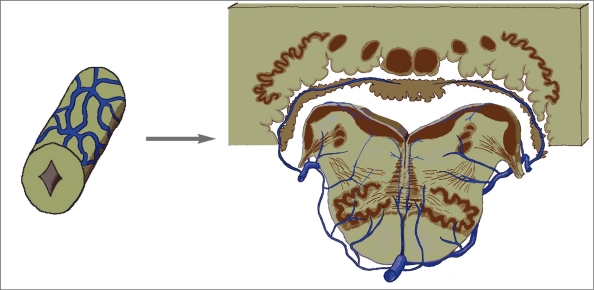
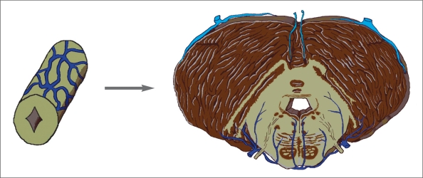
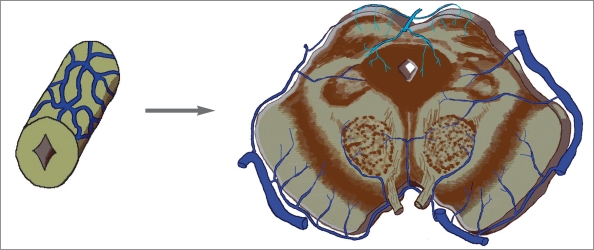
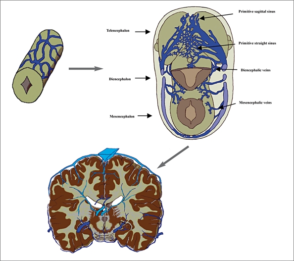
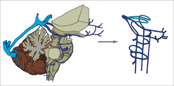
Ontogenetically, the ventricular venous systemmay develop in order to drain the gray matter(cells of the mantle layer of the neural tube) which migrates dorsally. On primitive brain vesicles of submammals especially fish, amphibianand reptile, the ventricular venous system is the major venous collector located on the middorsal surface, in between the meningeal layers comparable to the subarachnoid space in mammals. The ventricular venous system functions as a major drainage system for the brain vesicles in these submammals but its role decreases when the other two venous systems develop. Concerning the route of venous exit from the brain vesicles, we found that it resembles the spinal cord but could not be found all the way along the brain vesicles.
Figures
Similar articles
-
Patterns of the Cranial Venous System from the Comparative Anatomy in Vertebrates. Part II.The Lateral-Ventral Venous System.Interv Neuroradiol. 2008 Mar 30;14(1):21-31. doi: 10.1177/159101990801400103. Epub 2008 May 12. Interv Neuroradiol. 2008. PMID: 20557782 Free PMC article.
-
Patterns of cranial venous system from the comparative anatomy in vertebrates. Part I, introduction and the dorsal venous system.Interv Neuroradiol. 2007 Dec;13(4):335-44. doi: 10.1177/159101990701300404. Epub 2008 Feb 1. Interv Neuroradiol. 2007. PMID: 20566102 Free PMC article.
-
Anatomy of cerebral veins and sinuses.Front Neurol Neurosci. 2008;23:4-15. doi: 10.1159/000111256. Front Neurol Neurosci. 2008. PMID: 18004050 Review.
-
On the spinal venous sinus of Alligator mississippiensis.Anat Rec (Hoboken). 2024 Sep;307(9):2953-2965. doi: 10.1002/ar.25403. Epub 2024 Feb 7. Anat Rec (Hoboken). 2024. PMID: 38323749
-
Embryology of the Cavernous Sinus and Relevant Veins.J Neuroendovasc Ther. 2020;14(12):540-546. doi: 10.5797/jnet.ra.2020-0052. Epub 2020 Jun 19. J Neuroendovasc Ther. 2020. PMID: 37502138 Free PMC article. Review.
Cited by
-
Embryological Consideration of Dural AVFs in Relation to the Neural Crest and the Mesoderm.Neurointervention. 2019 Mar;14(1):9-16. doi: 10.5469/neuroint.2018.01095. Epub 2019 Feb 28. Neurointervention. 2019. PMID: 30827062 Free PMC article.
-
Embryological Consideration of Dural Arteriovenous Fistulas.Neurol Med Chir (Tokyo). 2016 Sep 15;56(9):544-51. doi: 10.2176/nmc.oa.2015-0313. Epub 2016 Jun 1. Neurol Med Chir (Tokyo). 2016. PMID: 27250699 Free PMC article.
References
-
- Cecon S, Minnich B, Lametschwandtner A. Vascularization of the Brains of the Atlantic and Pacific Hagfishes, Myxine glutinosa and Eptatretus stouti: A Scanning Electron Microscope Study of Vascular Corrosion Casts. J Morphol. 2002;253:51–63. - PubMed
-
- Roofe PG. The Endocranial Blood Vessels of Amblystoma Tigerinum. J Comp Neurol. 1934;61(2):257–293.
-
- Majewska-Michalska E. Vascularization of the brain in guinea pig. I: Gross anatomy of the arteries and veins. Folia Morphol (Warsz) 1994;53(4):249–268. - PubMed
-
- Armstrong LD, Horowitz A. The Brain Venous System of the Dog. Am J Anat. 1971;132(4):479–490. - PubMed
LinkOut - more resources
Full Text Sources
