

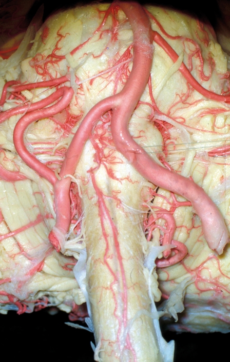
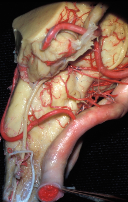
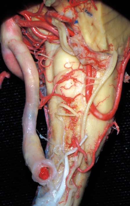
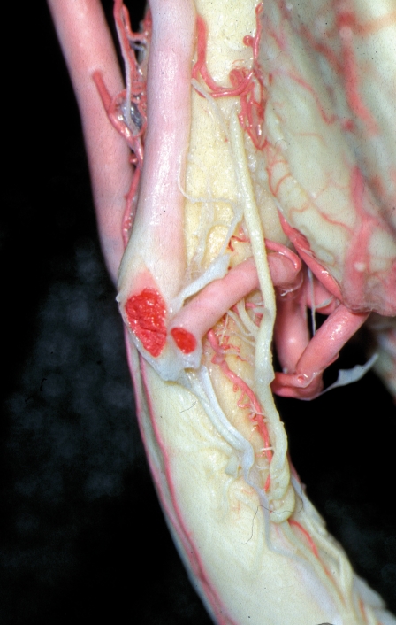
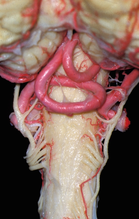
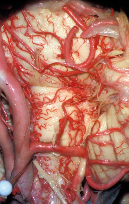
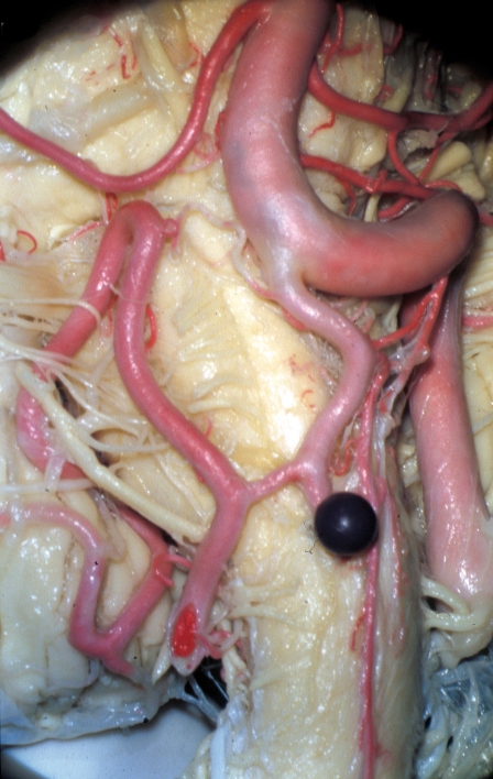
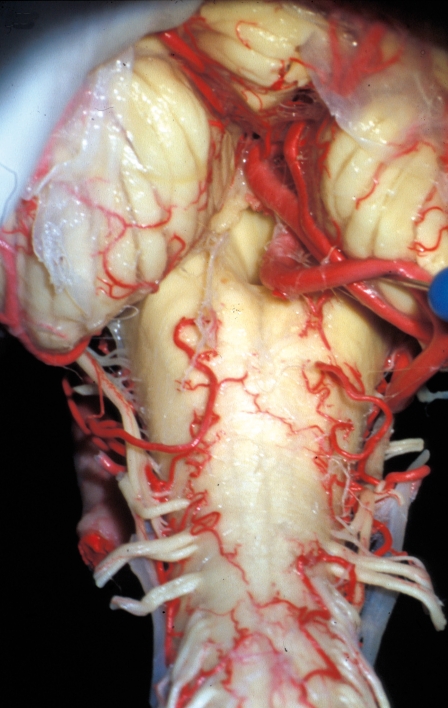
Vascular microanatomy of the pontomedullary junction, posterior inferior cerebellar arteries, and the lateral spinal arteries
- PMID: 20557786
- PMCID: PMC3313705
- DOI: 10.1177/159101990801400107
Vascular microanatomy of the pontomedullary junction, posterior inferior cerebellar arteries, and the lateral spinal arteries
Abstract
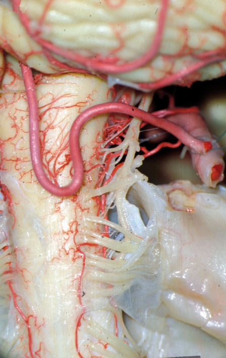
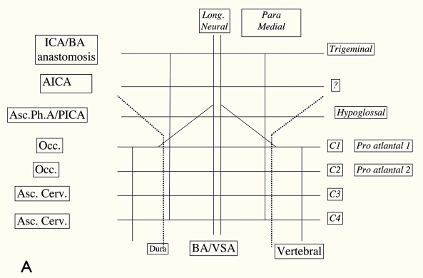
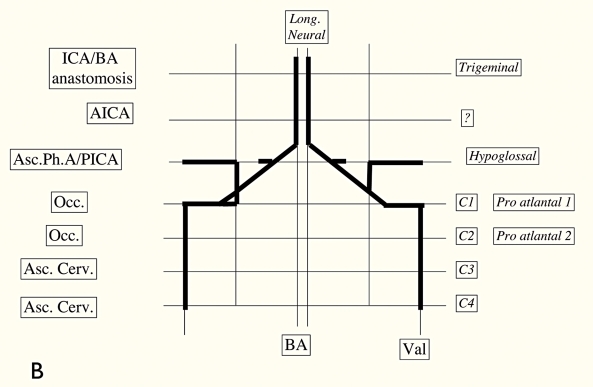
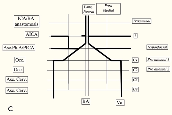
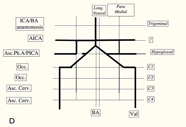
This study of 25 brains at the pontomedullary junction defined the different possible origins of the perforating arteries and lateral spinal arteries in relation to the posterior inferior cerebellar arteries (PICAs). - If the PICA emerges from the common trunk of the AICA-PICA coming from the basilar artery, it never gives perforating arteries or a lateral spinal artery on the lateral surface of the brain stem but supplies blood to a part of the ipsilateral cerebellar hemisphere. - If the PICA arises extradurally at C1, it never gives perforating arteries for the lateral surface of the brain stem, but it gives pial branches for the posterior surface of the medulla oblongata and is always the origin of the lateral spinal artery. - If the PICA emerges in the intradural vertebral artery, it is the source of the perforating arteries for the lateral surface of the brain stem and of the blood supply of the ipsilateral cerebellum.
Figures
References
-
- Akar ZC, Dujovny M, et al. Microvascular anatomy of the anterior surface of the medulla oblongata and olive. J Neurosurg. 1995;82:97–105. - PubMed
-
- Amarenco P, Hauw JJ. Anatomie des artères cérébelleuses. Rev Neurol. (Paris) 1989;145(4):267–276. - PubMed
-
- Ariens Kappers CU. Anatomie comparée du système nerveux. Paris: Masson et Cie; 1947. pp. 524–532.
-
- Duvernoy HM. Human Brainstem Vessels. Berlin Heidelberg New York: Springer-Verlag; 1978.
-
- Grand W, Budny JL, et al. Microvascular surgical anatomy of the vertebrobasilar junction. Neurosurg. 1997;40(6):1219–1225. - PubMed
LinkOut - more resources
Full Text Sources
