Management of posterior fossa dissecting aneurysms
- PMID: 20557803
- PMCID: PMC3328073
- DOI: 10.1177/15910199080140s212
Management of posterior fossa dissecting aneurysms
Abstract
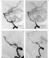
Treatment and prognosis of 14 patients of posterior fossa arterial dissections (AD) and dissecting aneurysms (DA) in one institution was reviewed. Internal trapping of aneurysm was performed for six patients presenting with SAH (three Vertebral, one posterior cerebral, one posterior inferior cerebellar, one anterior inferior cerebellar DA). The patency of the parent arteries was preserved in four DA patients with SAH (two Vertebral, two Basilar DA), 1 incidental vertebral DA, and one DA patient with brainstem infarction using stents and coils (four patients) or coils only (two patient). Proximal occlusion of parent artery was performed in a vertebral DA with SAH. One patient with a superior cerebellar DA presented with a midbrain infarct developed SAH with spontaneous occlusion of the aneurysm two weeks later. Of the 14 cases, ten were angiographically stable or cured during a follow up period of four to 70 months. one spontaneously resolved and two recurred. There was one death.
Figures


Similar articles
-
Dissecting aneurysms of the vertebral artery: a management strategy.J Neurosurg. 2002 Aug;97(2):259-67. doi: 10.3171/jns.2002.97.2.0259. J Neurosurg. 2002. PMID: 12186451
-
Predictive factors of medullary infarction after endovascular internal trapping using coils for vertebral artery dissecting aneurysms.J Neurosurg. 2018 Jul;129(1):107-113. doi: 10.3171/2017.2.JNS162916. Epub 2017 Aug 11. J Neurosurg. 2018. PMID: 28799869
-
[Endovascular management and classification of the dissecting aneurysms of the vertebral artery].Zhonghua Yi Xue Za Zhi. 2017 Jun 20;97(23):1773-1777. doi: 10.3760/cma.j.issn.0376-2491.2017.23.004. Zhonghua Yi Xue Za Zhi. 2017. PMID: 28647997 Chinese.
-
[Rerupture mechanism of ruptured intracranial dissecting aneurysm in the vertebral artery following proximal occlusion].No Shinkei Geka. 2000 Apr;28(4):345-9. No Shinkei Geka. 2000. PMID: 10769833 Review. Japanese.
-
Subarachnoid hemorrhage from vertebral artery dissecting aneurysms involving the origin of the posteroinferior cerebellar artery: report of two cases and review of the literature.Neurosurgery. 2000 Jan;46(1):196-200; discussion 200-1. Neurosurgery. 2000. PMID: 10626950 Review.
Cited by
-
Endovascular treatment of isolated posterior inferior cerebellar artery dissecting aneurysms: parent artery occlusion or selective coiling?Clin Neuroradiol. 2014 Sep;24(3):255-61. doi: 10.1007/s00062-013-0247-5. Epub 2013 Aug 4. Clin Neuroradiol. 2014. PMID: 23913020 Clinical Trial.
References
-
- Kitanaka C, Tanaki J, et al. Nonsurgical treatment of unruptured intracranial vertebral artery dissection with serial follow-up angiography. J Neurosurg. 1994;80:667–674. - PubMed
-
- Pozzati E, Padovani R, et al. Benign arterial dissections of the posterior circulation. J Neurosurg. 1991;75:69–72. - PubMed
-
- Maillo A, Díaz P, Morales F. Dissecting aneurysm of the posterior cerebral artery: spontaneous resolution. Neurosurgery. 1991;29:291–4294. - PubMed
LinkOut - more resources
Full Text Sources

