. 2010 Aug 12:1348:42-54.
doi: 10.1016/j.brainres.2010.06.021. Epub 2010 Jun 15.

Cloning and distribution of a putative octopamine/tyramine receptor in the central nervous system of the freshwater prawn Macrobrachium rosenbergii

Affiliations

Cloning and distribution of a putative octopamine/tyramine receptor in the central nervous system of the freshwater prawn Macrobrachium rosenbergii

Dalynés Reyes-Colón et al. Brain Res. .

Abstract

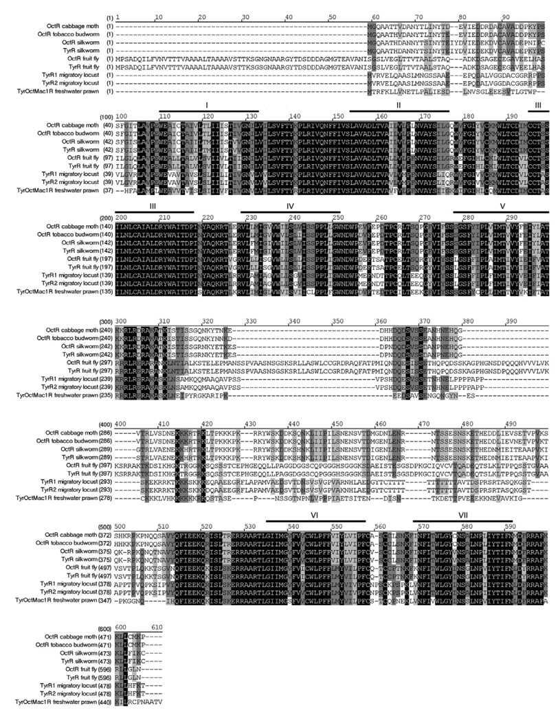
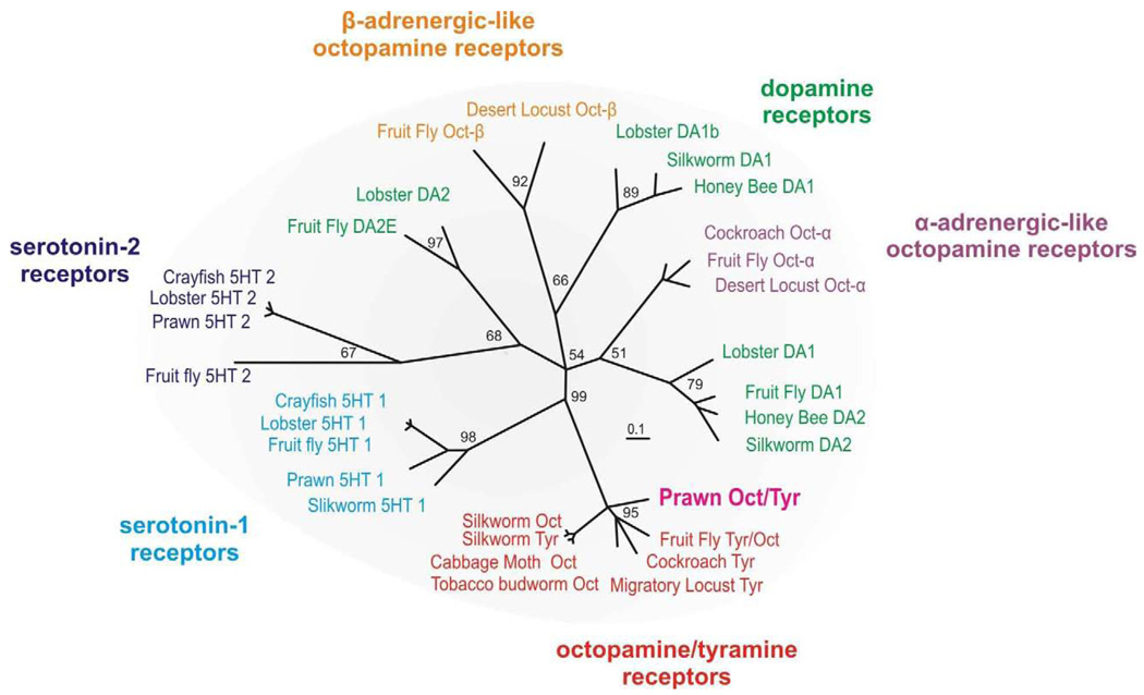
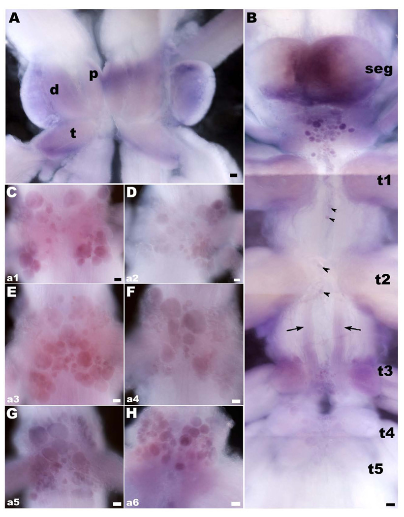
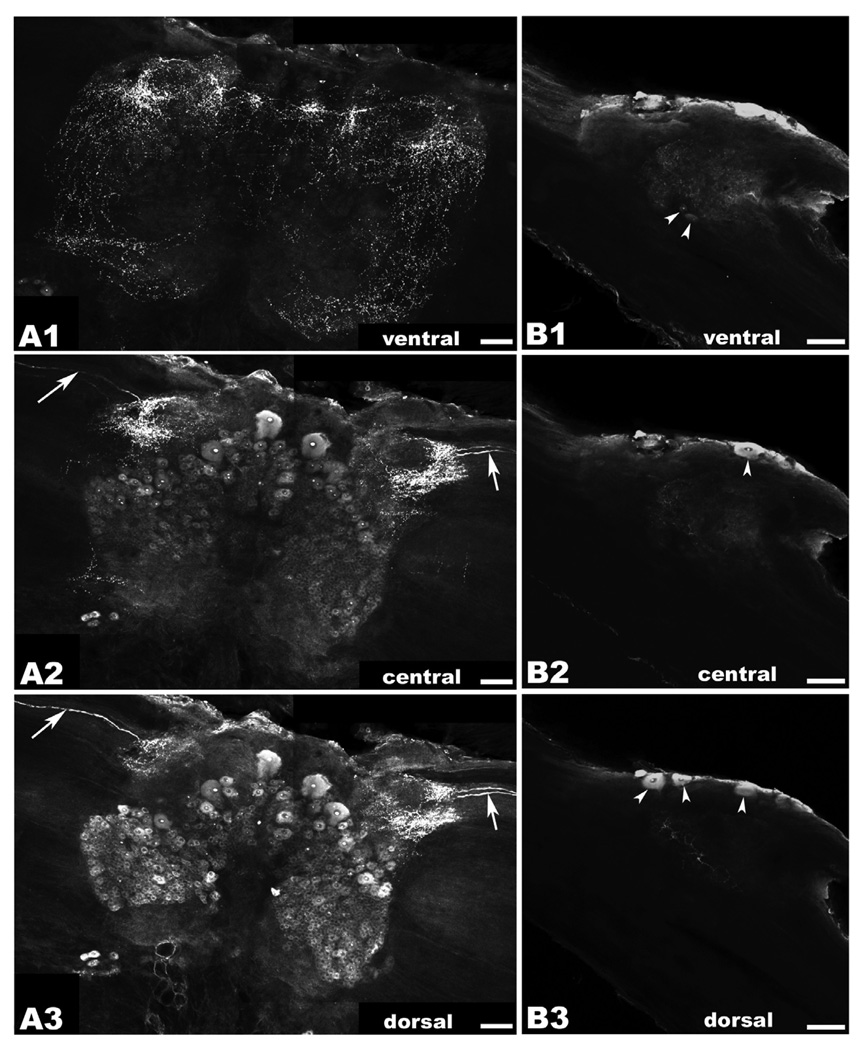
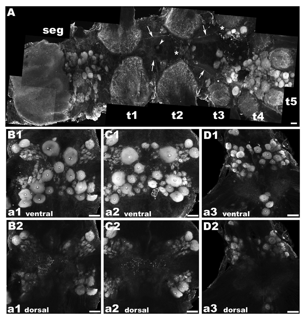
There is ample evidence linking octopamine (OA) and tyramine (TA) to several neurophysiological functions in arthropods. In our laboratory we use the freshwater prawn Macrobrachium rosenbergii to study the neural basis of aggressive behavior. As a first step towards understanding the possible role of these amines and their receptors in the modulation of interactive behaviors, we have cloned a putative octopamine/tyramine receptor. The predicted sequence of the cloned OA/TA(Mac) receptor consists of 1,579 base pairs (bp), with an open reading frame of 1,350bp that encodes a 450 amino acid protein. This putative protein displays sequence identities of 70% to an Aedes aegypti mosquito TA receptor, followed by 60% to a Stegomyia aegypti mosquito OA receptor, 59% and 58% to the migratory locust TA-1 and -2 receptors respectively, and 57% with the silkworm OA receptor. We also mapped the OA/TA(Mac) receptor distribution by in-situ hybridization to the receptor's mRNA, and by immunohistochemistry to its protein. We observed stained cell bodies for the receptor's mRNA, mainly in the midline region of the thoracic and in the abdominal ganglia, as well as diffuse staining in the brain ganglia. For the receptor's protein, we observed extensive punctate staining within the neuropil and on the membrane of specific groups of neurons in all ganglia throughout the CNS, including the brain, the midline region and neuropiles of the thoracic ganglia, and ventral part and neuropiles of the abdominal ganglia. The same pattern of stained cells was observed on the thoracic and abdominal ganglia in both in-situ hybridization and immunohistochemistry experiments. Diffuse staining observed with in-situ hybridization also coincides with punctate staining observed in brain, SEG, thoracic, and abdominal ganglia in immunohistochemical preparations. This work provides the first step towards characterizing the neural networks that mediate octopaminergic signaling in prawn.


Figures

Figure 1
Figure 1. Alignment of OA/TAMac receptor
The amino acid sequences of OA/TAMac are aligned with homologous invertebrate species. Heavy lines and Roman numbers above the sequence indicate transmembrane regions 1–7. Black boxes with white letters represent areas of identical regions, dark gray boxes with black letters represent conserved regions, and light gray boxes with black letters represent blocks of similar regions. Accession numbers are: cabbage moth Mamestra brasicae, AAK14402; tobacco budworm Heliothis virescens, Q25188; silkworm Bombyx mori, NP_001037504 and BAD11157; fly Drosophila melanogaster, AAA28731 and NP_524419; migratory locust LocusTYR Migratoria, Q25321 and Q25322; freshwater prawn Macrobrachium rosenbergii, EU233816. Abbreviations: octopamine receptor (OctR); tyramine receptor (TyrR); tyramine receptor 1 (TyrR1); tyramine receptor 2 (TyrR2); tyramine/octopamine receptor (Tyr/Oct).
Figure 2
Figure 2. Phylogenetic tree
The evolutionary relationship of the prawn’s putative OA/TAMac receptor to characterized biogenic amine receptors from other arthropod species indicates it belongs to the octopamine/tyramine family. See Experimental Procedure for the details of tree construction and sequence. Numbers at branches represent support values and the branch-length scale bar represents 0.1 amino acid substitutions per site.
Figure 3
Figure 3. OA/TAMac receptor’s mRNA in the CNS of the prawn
A: Brain OA/TAMac receptor mRNA appears as diffuse probe labeling in the neuropil areas of the protocerebrum (p), deutocerebrum (d), and tritocerebrum (t). B: Between the subesophageal ganglion (seg) and the first thoracic ganglia (t1), there is a central cluster of cell bodies of varying sizes that showed OA/TAMac mRNA staining. In the thoracic ganglia, two to four stained cell bodies showed mRNA labeling along the ventral midline (black arrowheads), as did a group of cells in the ventral middle region of the t3–t5 ganglia. There is also diffuse staining in a bilateral bundle of processes extending from t2 to t3 (black arrows). C–H: OA/TAMac mRNA Staining was also observed in all abdominal ganglia. Cells are arranged in clusters, following a pattern that repeated itself in each of the six ganglia (a1–a6). The pattern consisted of cells located ventrally at the center of each hemiganglion, along with 4 clusters of cells, arranged as the wings of a butterfly, located towards the dorsal lateral-most edges of each ganglion. Scale bar = 100 µm.
Figure 4
Figure 4. OA/TAMac immunoreactivity (ir) in the brain and circumesophageal (CEG) ganglia
A1–A3: Brain OA/TAMac-ir was observed as punctate staining in the ventral part of the ganglion (A1), in the protocerebrum, lateralmost aspects of the deutocerebrum, and extending towards the inferior aspects of the tritocerebrum. Moving more deeply towards the center of the ganglion (A2), punctate staining is observed in the lateralmost protocerebrum, while medium- and large-sized cells are found more medially. Highly immunoreactive structures within the nucleus, possibly nucleoli, are observed in all stained cells. Bilateral axons are observed in the optic nerves going towards a region of intense punctate staining in the lateral protocerebrum (white arrows). More dorsally (A3), protocerebrum punctate staining and the bilateral axons (white arrows) are still visible. Smaller-sized cell bodies showing OA/TAMac-ir are also observed in the deutocerebrum. B1–B3: In circumesophageal ganglia, OA/TAMac-ir was observed as punctate staining towards the center of the ventral (B1), middle (B2), and dorsal (B3) aspects of the ganglia. OA/TAMac-ir is also observed in two small cells in the ventral ganglia (white arrowheads; B1), and in one to three medium-sized cells on the outer surface of the ganglia (white arrowheads; B2–B3). Scale bar = 100 µm.
Figure 5
Figure 5. OA/TAMac immunoreactivity (ir) in the subesophageal (seg), thoracic (t), and abdominal (a) ganglia
A: OA/TAMac-ir was found in a central cluster of large cells in the inferior portion of the seg, superior to t1. Punctate staining was also found in the neuropil of all five thoracic ganglia. Single OA/TAMac-ir large cells were found in the midline of the ventral nerve cord, at the t1 and t2 levels (white arrowhead). The t3–t5 ganglia showed the most prominent staining in large centrally located clusters of multiple-sized cells. A pair of bilateral axons extending from t1 to t3 was observed (white arrows), crossing the ventral midline towards the contralateral side at the t2 level, and other of their ramifications going towards the central, lateral, and inferior parts of the ganglia at the t3 level (white asterisks). B–D: In the abdominal ganglia, staining was observed in cells arranged in clusters, following a “butterfly wing” pattern in the first five ganglia (a1–a5), while a6 also had a pair of clusters of large OA/TAMac-ir cells and four additional cells oriented longitudinally, central and inferior of the neuropil area, extending towards the nerve roots. A representation of the ventral part of a1 (B1), a2 (C1), and a6 (D1) is shown. Punctate OA/TAMac staining was also observed in the neuropil area of all six ganglia. A representation of the dorsal part of a1 (B2), a2 (C2), and a6 (D2) showing the neuropil region is shown. Scale bar = µm.

Similar articles

Cited by

References

    1. Antonsen BL, Paul DH. Serotonergic and octopaminergic systems in the squat lobster Munida quadrispina (Anomura, Glatheidae) J Comp Neurol. 2001;439:450–468. - PubMed
    1. Arakawa S, Gocayne JD, McCombie WR, Urquhart DA, Hall LM, Fraser CM, Vanter JC. Cloning, localization, and permanent expression of a Drosophila octopamine receptor. Neuron. 1990;4:343–354. - PubMed
    1. Axelrod J, Saavedra JM. Octopamine. Nature. 1977;265:501–504. - PubMed
    1. Barki A, Karplus I, Goren M. The agonistic behavior of the three male morphotypes of the freshwater prawn Macrobrachium rosenbergii (Crustacea, Palaemonidae) Behavior. 1991a;116:252–277.
    1. Barki A, Karplus I, Goren M. Morphotype related dominance hierarchies in males of Macrobrachium rosenbergii (Crustacea, Palaemoidae) Behavior. 1991b;117:145–160.

Publication types

MeSH terms