

Dynamic imaging of mammalian neural tube closure
- PMID: 20558153
- PMCID: PMC3873863
- DOI: 10.1016/j.ydbio.2010.06.010
Dynamic imaging of mammalian neural tube closure
Abstract
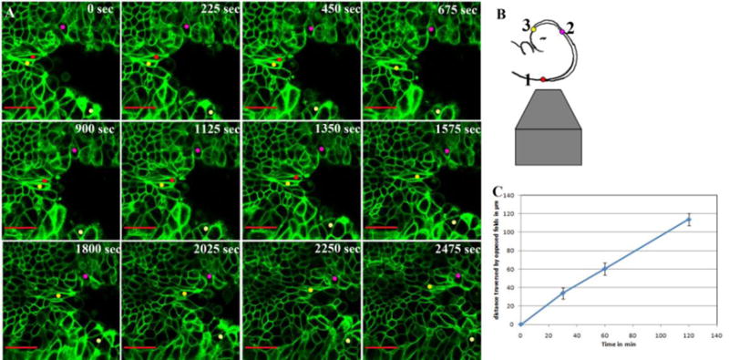
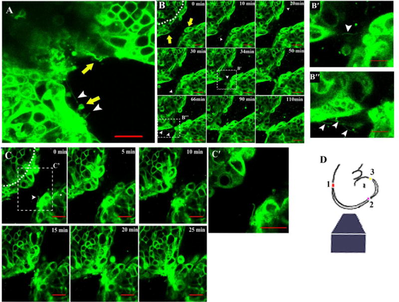
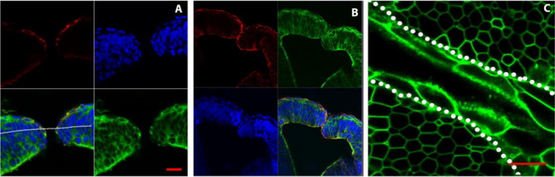
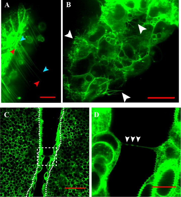
Neurulation, the process of neural tube formation, is a complex morphogenetic event. In the mammalian embryo, an understanding of the dynamic nature of neurulation has been hampered due to its in utero development. Here we use laser point scanning confocal microscopy of a membrane expressed fluorescent protein to visualize the dynamic cell behaviors comprising neural tube closure in the cultured mouse embryo. In particular, we have focused on the final step wherein the neural folds approach one another and seal to form the closed neural tube. Our unexpected findings reveal a mechanism of closure in the midbrain different from the zipper-like process thought to occur more generally. Individual non-neural ectoderm cells on opposing sides of the neural folds undergo a dramatic change in shape to protrude from the epithelial layer and then form intermediate closure points to "button-up" the folds. Cells from the juxtaposed neural folds extend long and short flexible extensions and form bridges across the physical gap of the closing folds. Thus, the combination of live embryo culture with dynamic imaging provides intriguing insight into the cell biological processes that mold embryonic tissues in mammals.
Copyright 2010 Elsevier Inc. All rights reserved.
Conflict of interest statement
The authors state there is no conflict of interest.
Figures
Similar articles
-
Integrin-Mediated Focal Anchorage Drives Epithelial Zippering during Mouse Neural Tube Closure.Dev Cell. 2020 Feb 10;52(3):321-334.e6. doi: 10.1016/j.devcel.2020.01.012. Dev Cell. 2020. PMID: 32049039 Free PMC article.
-
Apoptosis is not required for mammalian neural tube closure.Proc Natl Acad Sci U S A. 2009 May 19;106(20):8233-8. doi: 10.1073/pnas.0900333106. Epub 2009 May 6. Proc Natl Acad Sci U S A. 2009. PMID: 19420217 Free PMC article.
-
Regulation of cell protrusions by small GTPases during fusion of the neural folds.Elife. 2016 Apr 26;5:e13273. doi: 10.7554/eLife.13273. Elife. 2016. PMID: 27114066 Free PMC article.
-
Epithelial fusion during neural tube morphogenesis.Birth Defects Res A Clin Mol Teratol. 2012 Oct;94(10):817-23. doi: 10.1002/bdra.23072. Epub 2012 Sep 3. Birth Defects Res A Clin Mol Teratol. 2012. PMID: 22945349 Free PMC article. Review.
-
Morphogenetic movements in the neural plate and neural tube: mouse.Wiley Interdiscip Rev Dev Biol. 2014 Jan-Feb;3(1):59-68. doi: 10.1002/wdev.120. Epub 2013 May 29. Wiley Interdiscip Rev Dev Biol. 2014. PMID: 24902834 Review.
Cited by
-
Opportunities and Challenges in Tunneling Nanotubes Research: How Far from Clinical Application?Int J Mol Sci. 2021 Feb 25;22(5):2306. doi: 10.3390/ijms22052306. Int J Mol Sci. 2021. PMID: 33669068 Free PMC article. Review.
-
Role of Tunneling Nanotubes in the Nervous System.Int J Mol Sci. 2022 Oct 19;23(20):12545. doi: 10.3390/ijms232012545. Int J Mol Sci. 2022. PMID: 36293396 Free PMC article. Review.
-
Production of human spinal-cord organoids recapitulating neural-tube morphogenesis.Nat Biomed Eng. 2022 Apr;6(4):435-448. doi: 10.1038/s41551-022-00868-4. Epub 2022 Mar 28. Nat Biomed Eng. 2022. PMID: 35347276
-
Global analysis of cell behavior and protein dynamics reveals region-specific roles for Shroom3 and N-cadherin during neural tube closure.Elife. 2022 Mar 4;11:e66704. doi: 10.7554/eLife.66704. Elife. 2022. PMID: 35244026 Free PMC article.
-
Direct Cell-Cell Communication via Membrane Pores, Gap Junction Channels, and Tunneling Nanotubes: Medical Relevance of Mitochondrial Exchange.Int J Mol Sci. 2022 May 30;23(11):6133. doi: 10.3390/ijms23116133. Int J Mol Sci. 2022. PMID: 35682809 Free PMC article. Review.
References
-
- Bancroft M, Bellairs R. Differentiation of the neural plate and neural tube in the young chick embryo. A study by scanning and transmission electron microscopy. Anat Embryol (Berl) 1975;147:309–35. - PubMed
-
- Davidson LA, Keller RE. Neural tube closure in Xenopus laevis involves medial migration, directed protrusive activity, cell intercalation and convergent extension. Development. 1999;126:4547–56. - PubMed
-
- Fleming A, Gerrelli D, Greene ND, Copp AJ. Mechanisms of normal and abnormal neurulation: evidence from embryo culture studies. Int J Dev Biol. 1997;41:199–212. - PubMed
Publication types
MeSH terms
Grants and funding
LinkOut - more resources
Full Text Sources
Miscellaneous
