Comparative Study
. 2010 Aug 31;360(1-2):66-75.
doi: 10.1016/j.jim.2010.06.006. Epub 2010 Jun 15.

New monoclonal anti-mouse DC-SIGN antibodies reactive with acetone-fixed cells

Affiliations
Comparative Study

New monoclonal anti-mouse DC-SIGN antibodies reactive with acetone-fixed cells

Cheolho Cheong et al. J Immunol Methods. .

Abstract

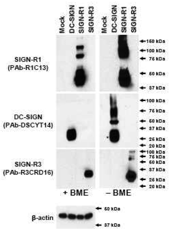
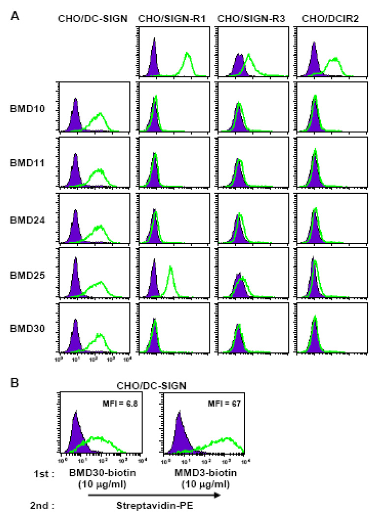
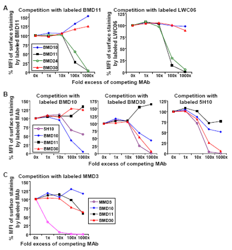
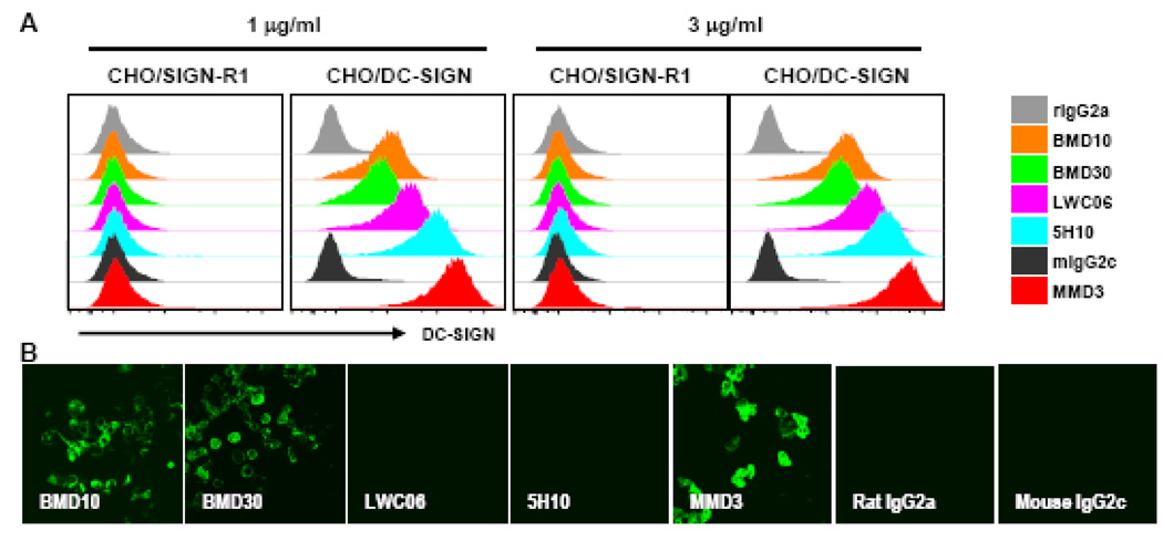
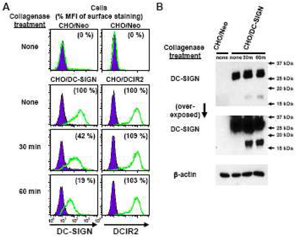
Mouse DC-SIGN CD209a is a type II transmembrane protein, one of a family of C-type lectin genes syntenic and homologous to human DC-SIGN. Current anti-mouse DC-SIGN monoclonal antibodies (MAbs) are unable to react with DC-SIGN in acetone-fixed cells, limiting the chance to visualize DC-SIGN in tissue sections. We first produced rabbit polyclonal PAb-DSCYT14 against a 14-aa peptide in the cytosolic domain of mouse DC-SIGN, and it specifically detected DC-SIGN and not the related lectins, SIGN-R1 and SIGN-R3 expressed in transfected CHO cells. MAbs were generated by immunizing rats and DC-SIGN knockout mice with the extracellular region of mouse DC-SIGN. Five rat IgG2a or IgM MAbs, named BMD10, 11, 24, 25, and 30, were selected and each MAb specifically detected DC-SIGN by FACS and Western blots, although BMD25 was cross-reactive to SIGN-R1. Two mouse IgG2c MAbs MMD2 and MMD3 interestingly bound mouse DC-SIGN but at 10 fold higher levels than the rat MAbs. When the binding epitopes of the new BMD and two other commercial rat anti-DC-SIGN MAbs, 5H10 and LWC06, were examined by competition assays, the epitopes of BMD11, 24, and LWC06 were identical or closely overlapping while BMD10, 30, and 5H10 were shown to bind different epitopes. MMD2 and MMD3 epitopes were on a 3rd noncompeting region of mouse DC-SIGN. DC-SIGN expressed on the cell surface was sensitive to collagenase treatment, as monitored by polyclonal and MAb. These new reagents should be helpful to probe the biology of DC-SIGN in vivo.


Figures

Figure 1
Figure 1. Specific detection of DC-SIGN by rabbit polyclonal antibody DSCYT14
CMV mammalian expression vectors containing no insert (Mock), mouse DC-SIGN, SIGN-R1, SIGN-R3 cDNA were transfected into 293TAg cells and cell lysates were prepared 1 day later. Equivalent amounts of cell lysates were boiled in sample buffers with (+) or without (−) β-mercaptoethanol (BME) prior to Western blot analyses with rabbit polyclonal antibodies against each mouse SIGN molecule or anti-β-actin antibody.
Figure 2
Figure 2. Specific binding of anti-DC-SIGN MAbs to stably transfected CHO cells
(A) CHO cells expressing three mouse SIGN molecules (DC-SIGN, SIGN-R1, and SIGN-R3) or DCIR2 were surface stained with the new anti-DC-SIGN MAbs BMD10, 11, 24, 25 and 30. BMD10, 11, 24, and 30 were detected by anti-rat IgG-PE/Cy5.5, while BMD25 was detected by anti-rat IgM-PE. The control CHO cells in the top row were stained by MAb 22D1 (anti-SIGN-R1), PAb-R3CRD16 (anti-SIGN-R3), and MAb 33D1 (anti-DCIR2) respectively. (B) CHO cells expressing mouse DC-SIGN were stained with an equal amount of biotinylated rat BMD30 or mouse MMD3 MAb to DC-SIGN followed by the detection of streptavidin-PE. Relative binding of BMD30 and MMD3 are shown as mean fluorescent index (MFI).
Figure 3
Figure 3. Competition assays for the binding of different anti-DC-SIGN MAbs
(A, B, C) CHO/DC-SIGN cells were pre-incubated for 10 min with indicated amounts of unlabeled competing MAbs prior to the incubation of each fluorescent-labeled anti-DC-SIGN MAb for 30 min. The binding of fluorescent-labeled MAb to CHO/DC-SIGN cells was calculated as the percent (%) value of each MFI (Median Fluorescence Intensity) compared to the MFI of CHO/DC-SIGN cells stained without competing MAbs.
Figure 4
Figure 4. Comparison of different anti-DC-SIGN MAbs for immunolabeling
(A)Anti-DC-SIGN MAbs that have been previously reported plus the new MAbs reported here were used to stain CHO/DC-SIGN or CHO/SIGN-R1 cells at 1 µg/ml and 3 µg/ml; labeling was assessed by FACS.(B) As in (A), but CHO/DC-SIGN cells grown on cover slides and fixed with acetone were stained. The binding was visualized with Streptavidin-APC in (A) and Streptavidin-Alexa 488 in (B).
Figure 5
Figure 5. Mouse DC-SIGN molecules are sensitive to collagenase
(A) CHO cells expressing mouse DC-SIGN, DCIR2, or control Neomycin (Neo) were incubated without (none) or with collagenase (400 units/ml) for 30 or 60 min, and the reaction was stopped by adding EDTA (10 mM final concentration). The halves of treated CHO cells were, then, stained with antibodies as indicated for FACS analysis. Relative levels of surface expression for each molecule were calculated as the percent (%) value of each MFI compared to the MFI of the CHO/DC-SIGN, or CHO/DCIR2 cells stained without collagenase treatment. (B) The remaining halves of CHO/Neo and CHO/DC-SIGN cells treated with/without collagenase from (A) were harvested and lysed. Equivalent amounts of cell lysates were boiled in sample buffer and separated in a 15 % SDS-PAGE gel prior to Western blot analyses with PAb-DSCYT14 and anti-β-actin antibody.

Similar articles

Cited by

References

    1. Alvarez CP, Lasala F, Carrillo J, Muniz O, Corbi AL, Delgado R. C-type lectins DC-SIGN and L-SIGN mediate cellular entry by Ebola virus in cis and in trans. J. Virol. 2002;76:6841–6844. - PMC - PubMed
    1. Banchereau J, Steinman RM. Dendritic cells and the control of immunity. Nature. 1998;392:245–252. - PubMed
    1. Boscardin SB, Hafalla JC, Masilamani RF, Kamphorst AO, Zebroski HA, Rai U, Morrot A, Zavala F, Steinman RM, Nussenzweig RS, Nussenzweig MC. Antigen targeting to dendritic cells elicits long-lived T cell help for antibody responses. J. Exp. Med. 2006;203:599–606. - PMC - PubMed
    1. Caminschi I, Corbett AJ, Zahra C, Lahoud M, Lucas KM, Sofi M, Vremec D, Gramberg T, Pohlmann S, Curtis J, Handman E, van Dommelen SL, Fleming P, Degli-Esposti MA, Shortman K, Wright MD. Functional comparison of mouse CIRE/mouse DC-SIGN and human DC-SIGN. Int. Immunol. 2006;18:741–753. - PMC - PubMed
    1. Caminschi I, Lucas KM, O'Keeffe MA, Hochrein H, Laabi Y, Brodnicki TC, Lew AM, Shortman K, Wright MD. Molecular cloning of a C-type lectin superfamily protein differentially expressed by CD8α− splenic dendritic cells. Mol. Immunol. 2001;38:365–373. - PubMed

Publication types

MeSH terms

LinkOut - more resources