

Histochemical detection of GM1 ganglioside using cholera toxin-B subunit. Evaluation of critical factors optimal for in situ detection with special emphasis to acetone pre-extraction
- PMID: 20558344
- PMCID: PMC3167299
- DOI: 10.4081/ejh.2010.e23
Histochemical detection of GM1 ganglioside using cholera toxin-B subunit. Evaluation of critical factors optimal for in situ detection with special emphasis to acetone pre-extraction
Abstract
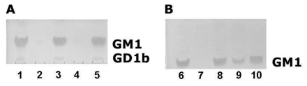
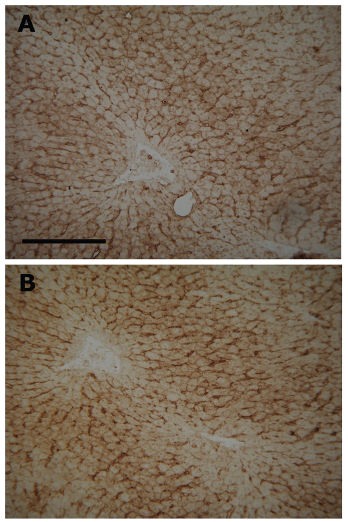
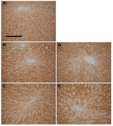
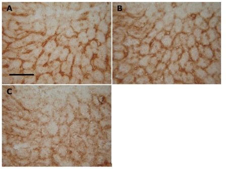
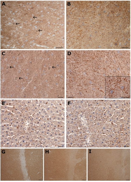
A comparison of histochemical detection of GM1 ganglioside in cryostat sections using cholera toxin B-subunit after fixation with 4% formaldehyde and dry acetone gave tissue-dependent results. In the liver no pre-treatment showed detectable differences related to GM1 reaction products, while studies in the brain showed the superiority of acetone pre-extraction (followed by formaldehyde), which yielded sharper images compared with the diffuse, blurred staining pattern associated with formaldehyde. Therefore, the aim of our study was to define the optimal conditions for the GM1 detection using cholera toxin B-subunit. Ganglioside extractability with acetone, the ever neglected topic, was tested comparing anhydrous acetone with acetone containing admixture of water. TLC analysis of acetone extractable GM1 ganglioside from liver sections did not exceed 2% of the total GM1 ganglioside content using anhydrous acetone at -20 degrees C, and 4% at room temperature. The loss increased to 30.5% using 9:1 acetone/water. Similarly, photometric analysis of lipid sialic acid, extracted from dried liver homogenates with anhydrous acetone, showed the loss of gangliosides into acetone 3.0 +/- 0.3% only. The loss from dried brain homogenate was 9.5 +/- 1.1%. Thus, anhydrous conditions (dry tissue samples and anhydrous acetone) are crucial factors for optimal in situ ganglioside detection using acetone pre-treatment. This ensures effective physical fixation, especially in tissues rich in polar lipids (precipitation, prevention of in situ diffusion), and removal of cholesterol, which can act as a hydrophobic blocking barrier.
Figures
Similar articles
-
Cholera toxin B subunit-binding and ganglioside GM1 immuno-expression are not necessarily correlated in human salivary glands.Acta Odontol Scand. 2014 Nov;72(8):694-700. doi: 10.3109/00016357.2014.898090. Epub 2014 Mar 21. Acta Odontol Scand. 2014. PMID: 24655314
-
Interleukin 3-dependent mouse mast cells express the cholera toxin-binding acidic glycosphingolipid, ganglioside GM1, and increase their histamine content in response to toxin.J Immunol. 1987 Sep 1;139(5):1640-6. J Immunol. 1987. PMID: 2957431
-
Cautionary note on the use of the B subunit of cholera toxin as a ganglioside GM1 probe: detection of cholera toxin A subunit in B subunit preparations by a sensitive adenylate cyclase assay.J Cell Biochem. 1990 Mar;42(3):143-52. doi: 10.1002/jcb.240420305. J Cell Biochem. 1990. PMID: 2156874
-
GM1 Ganglioside: Past Studies and Future Potential.Mol Neurobiol. 2016 Apr;53(3):1824-1842. doi: 10.1007/s12035-015-9136-z. Epub 2015 Mar 12. Mol Neurobiol. 2016. PMID: 25762012 Review.
-
Role of membrane gangliosides in the binding and action of bacterial toxins.J Membr Biol. 1982;69(2):85-97. doi: 10.1007/BF01872268. J Membr Biol. 1982. PMID: 6752418 Review.
Cited by
-
In situ detection of GM1 and GM2 gangliosides using immunohistochemical and immunofluorescent techniques for auxiliary diagnosis of canine and feline gangliosidoses.BMC Vet Res. 2016 Mar 31;12:67. doi: 10.1186/s12917-016-0691-y. BMC Vet Res. 2016. PMID: 27036194 Free PMC article.
-
Identifying pathological biomarkers: histochemistry still ranks high in the omics era.Eur J Histochem. 2011 Dec 7;55(4):e42. doi: 10.4081/ejh.2011.e42. Eur J Histochem. 2011. PMID: 22297448 Free PMC article.
-
Histochemistry through the years, browsing a long-established journal: novelties in traditional subjects.Eur J Histochem. 2010 Dec 16;54(4):e51. doi: 10.4081/ejh.2010.e51. Eur J Histochem. 2010. PMID: 21263750 Free PMC article.
-
δ-Opioid receptors: Pivotal role in intermittent hypoxia-augmentation of cardiac parasympathetic control and plasticity.Auton Neurosci. 2016 Jul;198:38-49. doi: 10.1016/j.autneu.2016.07.007. Epub 2016 Jul 25. Auton Neurosci. 2016. PMID: 27498137 Free PMC article.
-
On the future contents of a small journal of histochemistry.Eur J Histochem. 2012 Dec 10;56(4):e51. doi: 10.4081/ejh.2012.e51. Eur J Histochem. 2012. PMID: 23361247 Free PMC article.
References
-
- Hakomori S. Glycosphingolipids in cellular interaction differentiation and oncogenesis. In: Kanfer JN, Hakomori S, editors. Handbook of Lipid Research. Vol.3. Sphingolipid Biochemistry. Plenum Press; New York, London: 1983. pp. 327–336.
-
- Hakomori S. Glycosynapses: microdomains controlling carbohydrate-dependent cell adhesion and signaling. An Acad Bras Cienc. 2004;76:553–72. - PubMed
-
- Sonnino S, Mauri L, Chigorno V, Prinetti A. Gangliosides as components of lipid membrane domains. Glycobiology. 2007;17:1R–13R. - PubMed
-
- Holmgren J. Receptors for cholera toxin and Escherichia coli heat-labile enterotoxin revisited. Prog Brain Res. 1994;101:163–77. - PubMed
Publication types
MeSH terms
Substances
LinkOut - more resources
Full Text Sources
Other Literature Sources
