

Mitochondrial aldehyde dehydrogenase and cardiac diseases
- PMID: 20558439
- PMCID: PMC2936126
- DOI: 10.1093/cvr/cvq192
Mitochondrial aldehyde dehydrogenase and cardiac diseases
Abstract
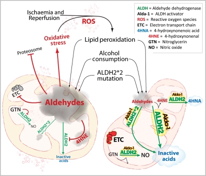
Numerous conditions promote oxidative stress, leading to the build-up of reactive aldehydes that cause cell damage and contribute to cardiac diseases. Aldehyde dehydrogenases (ALDHs) are important enzymes that eliminate toxic aldehydes by catalysing their oxidation to non-reactive acids. The review will discuss evidence indicating a role for a specific ALDH enzyme, the mitochondrial ALDH2, in combating oxidative stress by reducing the cellular 'aldehydic load'. Epidemiological studies in humans carrying an inactive ALDH2, genetic models in mice with altered ALDH2 levels, and small molecule activators of ALDH2 all highlight the role of ALDH2 in cardioprotection and suggest a promising new direction in cardiovascular research and the development of new treatments for cardiovascular diseases.
Figures
Similar articles
-
Metabolic remodeling induced by mitochondrial aldehyde stress stimulates tolerance to oxidative stress in the heart.Circ Res. 2009 Nov 20;105(11):1118-27. doi: 10.1161/CIRCRESAHA.109.206607. Epub 2009 Oct 8. Circ Res. 2009. PMID: 19815821
-
Mitochondrial aldehyde dehydrogenase 2 activation and cardioprotection.J Mol Cell Cardiol. 2013 Feb;55:58-63. doi: 10.1016/j.yjmcc.2012.03.017. Epub 2012 Apr 6. J Mol Cell Cardiol. 2013. PMID: 22507541 Review.
-
Mitochondrial aldehyde dehydrogenase 2 plays protective roles in heart failure after myocardial infarction via suppression of the cytosolic JNK/p53 pathway in mice.J Am Heart Assoc. 2014 Sep 18;3(5):e000779. doi: 10.1161/JAHA.113.000779. J Am Heart Assoc. 2014. PMID: 25237043 Free PMC article.
-
Mitochondrial Aldehyde Dehydrogenase in Myocardial Ischemic and Ischemia-Reperfusion Injury.Adv Exp Med Biol. 2019;1193:107-120. doi: 10.1007/978-981-13-6260-6_6. Adv Exp Med Biol. 2019. PMID: 31368100 Review.
-
Commentary: Aldehyde dehydrogenase, redox balance and exercise physiology: What is missing?Comp Biochem Physiol A Mol Integr Physiol. 2023 Sep;283:111470. doi: 10.1016/j.cbpa.2023.111470. Epub 2023 Jun 25. Comp Biochem Physiol A Mol Integr Physiol. 2023. PMID: 37364662
Cited by
-
Monoamine oxidase B prompts mitochondrial and cardiac dysfunction in pressure overloaded hearts.Antioxid Redox Signal. 2014 Jan 10;20(2):267-80. doi: 10.1089/ars.2012.4616. Epub 2013 May 22. Antioxid Redox Signal. 2014. PMID: 23581564 Free PMC article.
-
Mitochondrial aldehyde dehydrogenase (ALDH2) rescues cardiac contractile dysfunction in an APP/PS1 murine model of Alzheimer's disease via inhibition of ACSL4-dependent ferroptosis.Acta Pharmacol Sin. 2022 Jan;43(1):39-49. doi: 10.1038/s41401-021-00635-2. Epub 2021 Mar 25. Acta Pharmacol Sin. 2022. PMID: 33767380 Free PMC article.
-
Acetaldehyde dehydrogenase 2 interactions with LDLR and AMPK regulate foam cell formation.J Clin Invest. 2019 Jan 2;129(1):252-267. doi: 10.1172/JCI122064. Epub 2018 Dec 3. J Clin Invest. 2019. PMID: 30375985 Free PMC article.
-
Mitochondria as a source and target of lipid peroxidation products in healthy and diseased heart.Clin Exp Pharmacol Physiol. 2012 Feb;39(2):179-93. doi: 10.1111/j.1440-1681.2011.05641.x. Clin Exp Pharmacol Physiol. 2012. PMID: 22066679 Free PMC article. Review.
-
Transcriptomic Analysis of Trout Gill Ionocytes in Fresh Water and Sea Water Using Laser Capture Microdissection Combined with Microarray Analysis.PLoS One. 2015 Oct 6;10(10):e0139938. doi: 10.1371/journal.pone.0139938. eCollection 2015. PLoS One. 2015. PMID: 26439495 Free PMC article.
References
-
- Bolli R, Jeroudi MO, Patel BS, DuBose CM, Lai EK, Roberts R, et al. Direct evidence that oxygen-derived free radicals contribute to postischemic myocardial dysfunction in the intact dog. Proc Natl Acad Sci USA. 1989;86:4695–4699. doi:10.1073/pnas.86.12.4695. - DOI - PMC - PubMed
-
- Zhang M, Shah AM. Role of reactive oxygen species in myocardial remodeling. Curr Heart Fail Rep. 2007;4:26–30. doi:10.1007/s11897-007-0022-5. - DOI - PubMed
-
- Poyton RO, Ball KA, Castello PR. Mitochondrial generation of free radicals and hypoxic signaling. Trends Endocrinol Metab. 2009;20:332–340. - PubMed
-
- Turrens JF. Mitochondrial formation of reactive oxygen species. J Physiol. 2003;552:335–344. doi:10.1113/jphysiol.2003.049478. - DOI - PMC - PubMed
-
- Downey JM. Free radicals and their involvement during long-term myocardial ischemia and reperfusion. Annu Rev Physiol. 1990;52:487–504. doi:10.1146/annurev.ph.52.030190.002415. - DOI - PubMed
Publication types
MeSH terms
Substances
Grants and funding
LinkOut - more resources
Full Text Sources
Other Literature Sources
Medical
Miscellaneous
