. 2010 Nov 1;88(2):287-95.
doi: 10.1093/cvr/cvq193. Epub 2010 Jun 16.

Down's syndrome-like cardiac developmental defects in embryos of the transchromosomic Tc1 mouse

Affiliations

Down's syndrome-like cardiac developmental defects in embryos of the transchromosomic Tc1 mouse

Louisa Dunlevy et al. Cardiovasc Res. .

Abstract

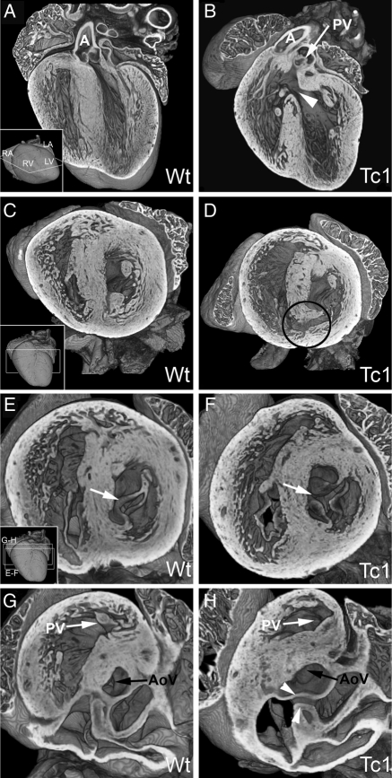
Aims: Cardiac malformations are prevalent in trisomies of human chromosome 21 [Down's syndrome (DS)], affecting normal chamber separation in the developing heart. Efforts to understand the aetiology of these defects have been severely hampered by the absence of an accurate mouse model. Such models have proved challenging to establish because synteny with human chromosome Hsa21 is distributed across three mouse chromosomes. None of those engineered so far accurately models the full range of DS cardiac phenotypes, in particular the profound disruptions resulting from atrioventricular septal defects (AVSDs). Here, we present analysis of the cardiac malformations exhibited by embryos of the transchromosomic mouse line Tc(Hsa21)1TybEmcf (Tc1) which contains more than 90% of chromosome Hsa21 in addition to the normal diploid mouse genome.

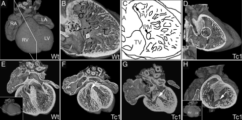
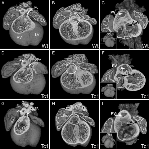
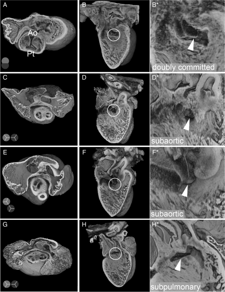
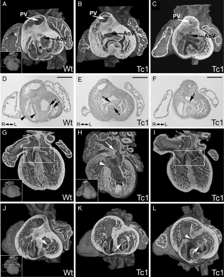
Methods and results: Using high-resolution episcopic microscopy and three-dimensional (3D) modelling, we show that Tc1 embryos exhibit many of the cardiac defects found in DS, including balanced AVSD with single and separate valvar orifices, membranous and muscular ventricular septal defects along with outflow tract and valve leaflet abnormalities. Frequencies of cardiac malformations (ranging from 38 to 55%) are dependent on strain background. In contrast, no comparable cardiac defects were detected in embryos of the more limited mouse trisomy model, Dp(16Cbr1-ORF9)1Rhr (Ts1Rhr), indicating that trisomy of the region syntenic to the Down's syndrome critical region, including the candidate genes DSCAM and DYRK1A, is insufficient to yield DS cardiac abnormalities.

Conclusion: The Tc1 mouse line provides a suitable model for studying the underlying genetic causes of the DS AVSD cardiac phenotype.


Figures

Figure 1
Figure 1
E14.5 Tc1 embryos exhibit ventricular septal defects. Three-dimensional reconstructions of wild-type (A, B, and E) and Tc1 (D, F, G, and H) hearts, eroded in the planes indicated in insets. (B and C) Division of the ventricular septum (right view) into membranous (M), outlet (O), inlet (I), and trabeculated (T) regions. Many Tc1 samples have defects in the membranous region (white circle, D and inset). (EG) Four-chambered view showing membranous VSD (white arrow, F) and muscular inlet VSD (white arrow, G; the defect has an exclusively muscular border and opens to the inlet region of the ventricular septum) in Tc1 hearts. (H) Short-axis view of a Tc1 muscular inlet VSD (white arrow, H). (LA, left atrium; LV, left ventricle; RA, right atrium; RV, right ventricle; M, membranous region; O, outlet region; I, inlet region; T, trabeculated region; A, aorta; PV, pulmonary valve; PM, base of papillary muscle; TV, septal leaflet of tricuspid valve).
Figure 2
Figure 2
E14.5 Tc1 embryos exhibit outflow tract defects. Three-dimensional reconstructions of wild-type (A–C) and Tc1 (DF, GI) hearts. In all samples analysed, the pulmonary artery (PA, white arrow, A, D, and G) arose from the right ventricle (RV). In the wild-type, the aorta arises from the left ventricle (B and C), whereas in the Tc1 hearts, the position of the aortic valve (white circle, B, E, and H) is shifted rightwards. Tc1 embryos can exhibit DORV, both the pulmonary artery and the aorta arising from the right ventricle (E and F) and overriding aorta, the aortic valve (AV) sitting centrally above a VSD, resulting in subaortic interventricular communication (H and I). The models in C, F, and I have been eroded to the level of the white box indicated in the respective insets. (LA, left atrium; LV, left ventricle; RA, right atrium; RV, right ventricle; PA, pulmonary artery; A, aorta; PV, pulmonary valve; AoV, aortic valve).
Figure 3
Figure 3
Abnormal arterial trunk arrangements in E14.5 Tc1 embryos. Three-dimensional reconstructions of Tc1 hearts, showing abnormal arterial trunk arrangements associated with DORV. Models are eroded in the short axis through the atria at the level of the aortic valve (A, C, E, and G) and the relative position of the aortic and pulmonary trunks is shown in diagram (pale and dark grey, respectively). For each heart, a long-axis view of the interventricular septum from the right ventricle (B, D, F, and H) reveals the location of the septal defect (white circle), shown in detail in the adjacent panel (B*, D*, F*, and H*; white arrowhead). Abnormalities range from failure of outflow tract septation, resulting in aortic-pulmonary continuity, with a doubly committed VSD (A and B); subaortic ventricular communication with bilateral infundibulums and either side-by-side (C and D) or normal arrangement of arterial trunks (E and F); subpulmonary VSD with side-by-side arterial trunks, equivalent to the Taussig–Bing anomaly in humans (G and H). (Ao, aortic trunk; Pt, pulmonary trunk).
Figure 4
Figure 4
E14.5 Tc1 embryos exhibit AV canal defects. Three-dimensional reconstructions of wild-type (A, G, and J) and Tc1 (B, C, H, I, K, and L) hearts eroded in the short or long axis (as indicated). (DF) The corresponding sections (haematoxylin and eosin stain) from (AC). In wild-type hearts, the two AV junctions are separated by the AV septum (black asterix, A) and the left outflow tract (black arrow, A) sits in a ‘wedged’ position between the mitral valve and AV septum. The tricuspid and mitral valves (D: black arrowheads and arrows, respectively) are therefore separate. In Tc1 hearts with AVSD (B, C, E, and F), a common AV junction can be guarded by a single valve (B) containing bridging leaflets (black arrows, E), or the bridging leaflets can be joined along the crest of the ventricular septum (black arrow, F), giving two valvar orifices within the common AV junction (C). In both cases, the ‘unwedged’ position of the left outflow tract is evident (black arrows, B and C). The AV bridging leaflets in Tc1 samples with AVSD are anchored to the atrial septum (white arrow, H) but not the ventricular septum (white arrowhead, H). Other Tc1 samples show fat enlarged valves within the AV junctions (compare valve leaflets in white boxes, G and I; leaflet thickness marked) or a cleft (or failure of fusion) in the anterior leaflet of the mitral valve (white arrows, K and L) either with (white arrowhead, L) or without (K) a VSD. (PV, pulmonary valve; AoV, aortic valve; R, right; L, left. Scale bars in D, E, and F represent 500 µm.)
Figure 5
Figure 5
E18.5 Tc1 embryos exhibit AVSD, ventricular septal, and outflow tract defects. Three-dimensional reconstructions of wild-type (A, C, E, and G) and Tc1 (B, D, F, and H) hearts eroded to the levels indicated in the insets. (B) A Tc1 membranous VSD (white arrowhead; four-chamber view) resulting in subpulmonary interventricular communication, both arterial roots arising side-by-side from the right ventricle (the Taussig–Bing malformation). (CH) Short-axis view from apex. (D) A Tc1 muscular inlet VSD (black circle, D). The left AV valve in the Tc1 heart is trifoliate in comparison to the normal mitral valve (compare E and F, white arrows). This apparent cleft in the aortic leaflet can be traced in continuity with the two bridging leaflets (white arrowheads, H) guarding a common AV junction (see also Supplementary material online, Movie S9) (AoV, aortic valve; LA, left atrium; LV, left ventricle; RA, right atrium; RV, right ventricle; A, aorta; PV, pulmonary valve; R, right; L, left; VS, ventricular septum).

Comment in

  • Looking down the atrioventricular canal.
    Benson DW, Sund KL. Benson DW, et al. Cardiovasc Res. 2010 Nov 1;88(2):205-6. doi: 10.1093/cvr/cvq302. Epub 2010 Sep 20. Cardiovasc Res. 2010. PMID: 20855523 Free PMC article. No abstract available.

Similar articles

Cited by

References

    1. Sherman SL, Allen EG, Bean LH, Freeman SB. Epidemiology of Down syndrome. Ment Retard Dev Disabil Res Rev. 2007;13:221–227. doi:10.1002/mrdd.20157. - DOI - PubMed
    1. Freeman SB, Taft LF, Dooley KJ, Allran K, Sherman SL, Hassold TJ, et al. Population-based study of congenital heart defects in Down syndrome. Am J Med Genet. 1998;80:213–217. doi:10.1002/(SICI)1096-8628(19981116)80:3<213::AID-AJMG6>3.0.CO;2-8. - DOI - PubMed
    1. Korenberg JR, Chen XN, Schipper R, Sun Z, Gonsky R, Gerwehr S, et al. Down syndrome phenotypes: the consequences of chromosomal imbalance. Proc Natl Acad Sci USA. 1994;91:4997–5001. doi:10.1073/pnas.91.11.4997. - DOI - PMC - PubMed
    1. Delabar JM, Theophile D, Rahmani Z, Chettouh Z, Blouin JL, Prieur M, et al. Molecular mapping of twenty-four features of Down syndrome on chromosome 21. Eur J Hum Genet. 1993;1:114–124. - PubMed
    1. Korbel JO, Tirosh-Wagner T, Urban AE, Chen XN, Kasowski M, Dai L, et al. The genetic architecture of Down syndrome phenotypes revealed by high-resolution analysis of human segmental trisomies. Proc Natl Acad Sci USA. 2009;106:12031–12036. doi:10.1073/pnas.0813248106. - DOI - PMC - PubMed

Publication types

MeSH terms