. 2010 Nov 1;88(2):324-33.
doi: 10.1093/cvr/cvq190. Epub 2010 Jun 16.

Consecutive pharmacological activation of PKA and PKC mimics the potent cardioprotection of temperature preconditioning

Affiliations

Consecutive pharmacological activation of PKA and PKC mimics the potent cardioprotection of temperature preconditioning

Igor Khaliulin et al. Cardiovasc Res. .

Abstract

Aims: Temperature preconditioning (TP) provides very powerful protection against ischaemia/reperfusion. Understanding the signalling pathways involved may enable the development of effective pharmacological cardioprotection. We investigated the interrelationship between activation of protein kinase A (PKA) and protein kinase C (PKC) in the signalling mechanisms of TP and developed a potent pharmacological intervention based on this mechanism.

Methods and results: Isolated rat hearts were subjected to TP, 30 min global ischaemia, and 60 min reperfusion. Other control and TP hearts were perfused with either sotalol (β-adrenergic blocker) or H-89 (PKA inhibitor). Some hearts were pre-treated with either isoproterenol (β-adrenergic agonist) or adenosine (PKC activator) that were given alone, simultaneously, or sequentially. Pre-treatment with isoproterenol, adenosine, and the consecutive isoproterenol/adenosine treatment was also combined with the PKC inhibitor chelerythrine. Cardioprotection was evaluated by haemodynamic function recovery, lactate dehydrogenase release, measurement of mitochondrial permeability transition pore opening, and protein carbonylation during reperfusion. Cyclic AMP and PKA activity were increased in TP hearts. H-89 and sotalol blocked the cardioprotective effect of TP and TP-induced PKC activation. Isoproterenol, adenosine, and the consecutive treatment increased PKC activity during pre-ischaemia. Isoproterenol significantly reduced myocardial glycogen content. Isoproterenol and adenosine, alone or simultaneously, protected hearts but the consecutive treatment gave the highest protection. Cardioprotective effects of adenosine were completely blocked by chelerythrine but those of the consecutive treatment only attenuated.

Conclusion: The signal transduction pathway of TP involves PKA activation that precedes PKC activation. Pharmacologically induced consecutive PKA/PKC activation mimics TP and induces extremely potent cardioprotection.


Figures
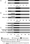
Figure 1
Figure 1
Outline of the protocols used in the experiments. (A) Series 1. TP, temperature preconditioning; C, control. (B) Series 2. CS and CH, control hearts perfused with 10 µM sotalol or H-89, respectively. TPS and TPH, TP hearts perfused with 10 µM sotalol or H-89, respectively. (C) Series 3 and 4. Iso, hearts perfused with 0.2 µM isoproterenol (2 min, 37°C) + 10 min washout; Ade, hearts perfused with 30 µM adenosine (5 min, 37°C) + 5 min washout; consecutive Iso + Ade, hearts perfused with isoproterenol then adenosine + 5 min washout; mixed Iso + Ade, 2 min perfusion with 0.2 µM isoproterenol during 5 min perfusion with 30 µM adenosine followed by 5 min washout. In Series 4, Iso, Ade, and the consecutive Iso + Ade treatments were also combined with 10 µM chelerythrine infusion started 5 min before perfusion with isoproterenol or adenosine and completed at the end of pre-ischaemia.
Figure 2
Figure 2
Effect of TP on PKA activity, cAMP concentration, and Akt/GSK3 phosphorylation. (A) PKA activity was measured using the PepTag® assay (Promega) and expressed as a ratio of fluorescence intensity of phosphorylated and non-phosphorylated PepTag® A1 peptide (P-A1 and A1, respectively). (Inset) A representative gel containing A1 and P-A1; Neg C, negative control; Pos C, positive control (PepTag® A1 peptide phosphorylated by the PKA catalytic subunit). (B) cAMP concentration was determined using a direct enzyme immunoassay kit (Sigma). (CF) GSK3 (C + E) and Akt (D + F) phosphorylation were determined prior to ischaemia (C + D) or after 15 min reperfusion (E + F) by western blotting using the ratio of band intensity of phosphorylated to total protein. Eight hearts each of control (C) and temperature preconditioning (TP) groups were used for all parameters. *P < 0.05 vs. control. Inset in each of (CF): representative blots of phosphorylated (P) and total (T) GSK3 and Akt.
Figure 3
Figure 3
Effect of sotalol and H-89 on RPP and PKC activity of control and TP hearts prior to ischaemia. (A) Mean data for RPP measured during hypothermic and normothermic perfusion in TP (n = 8), TP + 10 µM sotalol (TPS; n = 6), and TP + 10 µM H-89 (TPH; n = 6) hearts. RPP values for TPH and TPS groups were significantly lower (P < 0.05) than TP during all three episodes of normothermic perfusion. (B) Mean data for PKC activity measured in eight hearts each of control (C), TP, control + 10 µM H-89 (CH), and TPH groups. PKC activity was measured using non-radioactive PepTag® assay and is expressed as a ratio of fluorescence intensity of phosphorylated and non-phosphorylated peptide. *P < 0.05, **P < 0.01 vs. TP. (Inset) Representative gels containing non-phosphorylated and phosphorylated PepTag® C1 peptide (C1 and P-C1, respectively). Specificity of the PepTag® C1 peptide to PKC was confirmed by its reaction with PKC control enzyme (Pos C, positive control) and a heart sample (S). No phosphorylated peptide was found without the control enzyme (Neg C, negative control) or with the boiled heart sample (S-B).
Figure 4
Figure 4
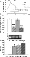
Effect of isoproterenol and adenosine on RPP, glycogen content, and PKC activity in the hearts prior to ischaemia. (A) Mean data for RPP in 0.2 µM isoproterenol (Iso; n = 7), 30 µM adenosine (Ade; n = 8), and consecutive isoproterenol + adenosine (C-Iso + Ade; n = 11) hearts measured during pre-ischaemia following the equilibration period. Isoproterenol significantly increased and adenosine reduced RPP compared with control hearts (C). The decrease in RPP was significantly greater in the C-Iso + Ade hearts than in the Ade hearts (P < 0.05) starting from 27 min pre-ischaemia. (B) Mean data for glycogen content in six hearts each of control C, Iso, Ade, and C-Iso + Ade groups. ***P < 0.001 vs. control. (C) Mean data for PKC activity in six each of C, Iso, Ade, and C-Iso + Ade groups measured in hearts prior to ischaemia. *P < 0.05 vs. control. (Inset) A representative gel containing non-phosphorylated and phosphorylated PepTag® C1 peptide (C1 and P-C1, respectively).
Figure 5
Figure 5
Effect of isoproterenol and adenosine on Ca2+-induced MPTP opening (A) and protein carbonylation (B). (A) Mean data for the rate of Ca2+-induced mitochondria swelling in C (n = 8), Iso (n = 7), Ade (n = 7), and C-Iso + Ade (n = 9) groups of hearts. Mitochondria were isolated after 30 min global ischaemia and MPTP opening determined under de-energized conditions as described under Section 2. *P < 0.05, **P < 0.01 vs. C, ##P < 0.01 vs. Iso + Ade. (B) Protein carbonylation measured by western blotting in mitochondria isolated from eight hearts each of non-ischaemic control (CP) and C, Iso, Ade, and C-Iso + Ade groups after 30 min global ischaemia. A representative blot is shown plus mean data for each condition. ND, non-derivatized control used to correct for non-specific binding. Further details are given in Supplementary Methods. **P< 0.01 vs. control.

References

    1. Depre C, Taegtmeyer H. Metabolic aspects of programmed cell survival and cell death in the heart. Cardiovasc Res. 2000;45:538–548. doi:10.1016/S0008-6363(99)00266-7. - DOI - PubMed
    1. Khaliulin I, Clarke SJ, Lin H, Parker JE, Suleiman MS, Halestrap AP. Temperature preconditioning of isolated rat hearts—a potent cardioprotective mechanism involving a reduction in oxidative stress and inhibition of the mitochondrial permeability transition pore. J Physiol. 2007;581:1147–1161. doi:10.1113/jphysiol.2007.130369. - DOI - PMC - PubMed
    1. Clarke SJ, Khaliulin I, Das M, Parker JE, Heesom KJ, Halestrap AP. Inhibition of mitochondrial permeability transition pore opening by ischemic preconditioning is probably mediated by reduction of oxidative stress rather than mitochondrial protein phosphorylation. Circ Res. 2008;102:1082–1090. doi:10.1161/CIRCRESAHA.107.167072. - DOI - PMC - PubMed
    1. Lochner A, Genade S, Tromp E, Podzuweit T, Moolman JA. Ischemic preconditioning and the β-adrenergic signal transduction pathway. Circulation. 1999;100:958–966. - PubMed
    1. Yellon DM, Dana A. The preconditioning phenomenon: a tool for the scientist or a clinical reality? Circ Res. 2000;87:543–550. - PubMed

Publication types

MeSH terms