Review
. 2010 Oct;51(10):2863-95.
doi: 10.1194/jlr.R005959. Epub 2010 Jun 17.

Biochemistry and genetics of inherited disorders of peroxisomal fatty acid metabolism

Affiliations
Review

Biochemistry and genetics of inherited disorders of peroxisomal fatty acid metabolism

Paul P Van Veldhoven. J Lipid Res. 2010 Oct.

Abstract

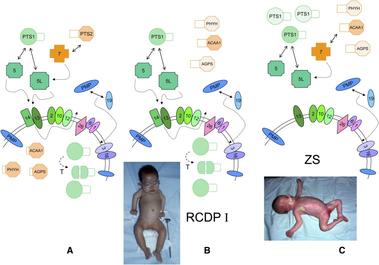
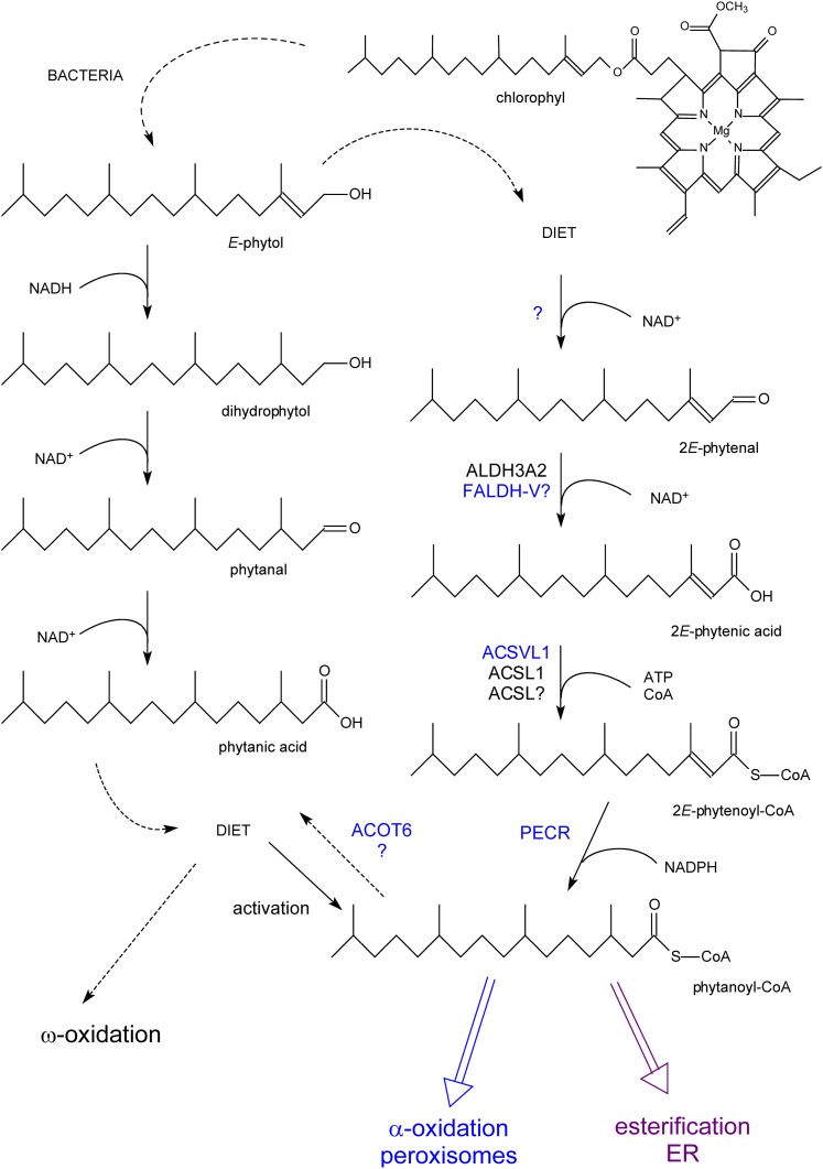
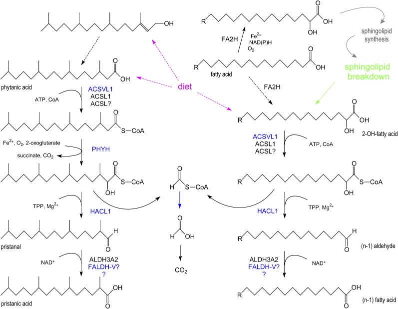
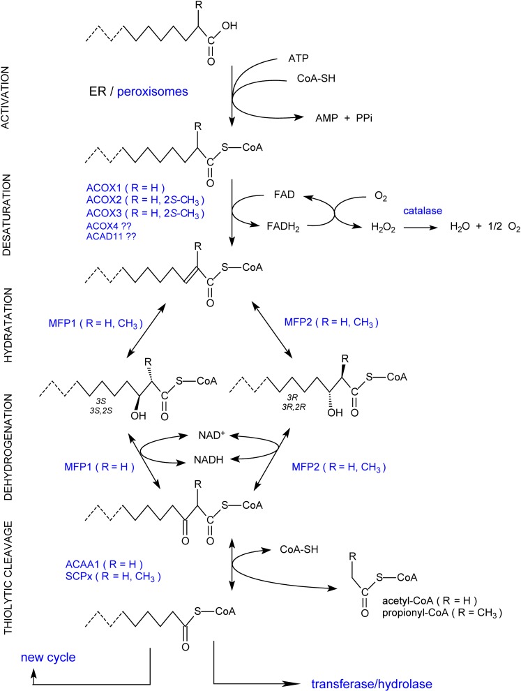
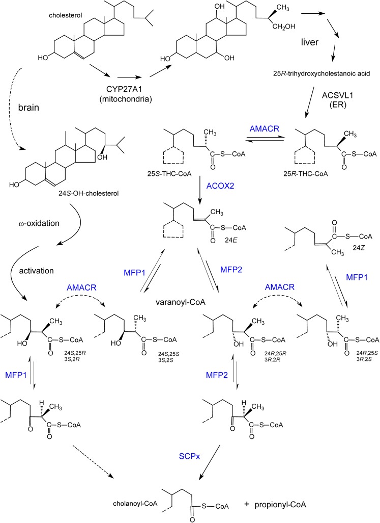
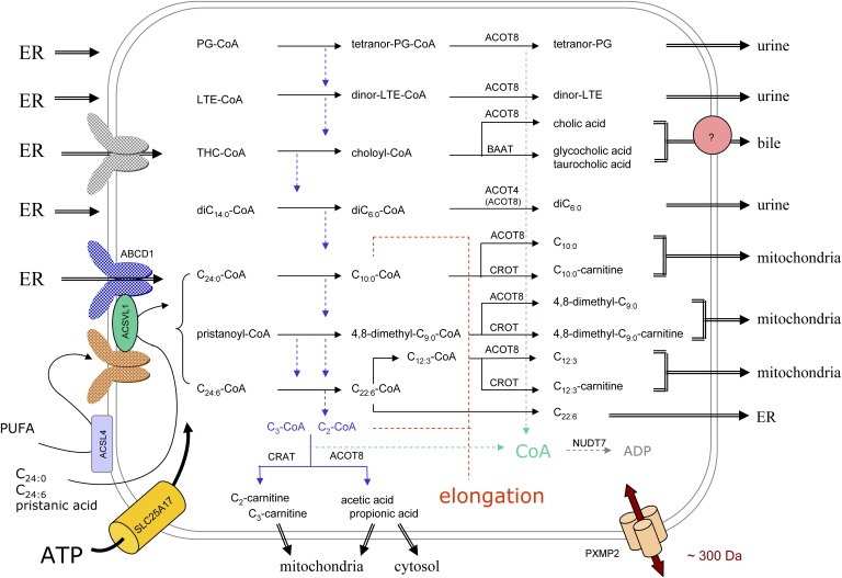
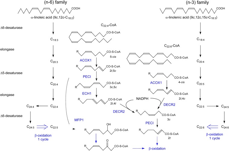
In humans, peroxisomes harbor a complex set of enzymes acting on various lipophilic carboxylic acids, organized in two basic pathways, alpha-oxidation and beta-oxidation; the latter pathway can also handle omega-oxidized compounds. Some oxidation products are crucial to human health (primary bile acids and polyunsaturated FAs), whereas other substrates have to be degraded in order to avoid neuropathology at a later age (very long-chain FAs and xenobiotic phytanic acid and pristanic acid). Whereas total absence of peroxisomes is lethal, single peroxisomal protein deficiencies can present with a mild or severe phenotype and are more informative to understand the pathogenic factors. The currently known single protein deficiencies equal about one-fourth of the number of proteins involved in peroxisomal FA metabolism. The biochemical properties of these proteins are highlighted, followed by an overview of the known diseases.


Figures

Fig. 1.
Fig. 1.
Peroxisome biogenesis in humans. A: A simplified scheme for protein import in mammalian peroxisomes under normal conditions and involved peroxins, indicated by their PEX number and arranged according to known interactions, is shown. The complex of PTS1-matrix proteins (in green) and one of the PEX5 isoforms, formed in the cytoplasm, will dock at the membrane, followed by translocation involving the RING peroxins PEX2, 10, and 12 (dashed arrows). After cargo release, PEX5 is recycled to the cytoplasm with the help of PEX1, 6, and 26 (dashed arrow). The role of mono- and polyubiquitination of PEX5 in this process is not displayed. Import of PTS2-matrix proteins (in orange), captured by PEX7, is mediated by the long PEX5 isoform. After import, some PTS1 proteins are proteolytically processed (T), and the targeting signal of PTS2-proteins is removed (ACAA1, thiolase; PHYH, AGPS). Newly synthesized PMPs (in blue) can interact with PEX19 in the cytoplasm (or with the membrane associated PEX19), followed by docking and membrane insertion. Peroxins playing a role in PMP targeting are depicted in different shades of blue. In the absence of a functional PEX7, PTS2 proteins are not imported and processed and are degraded in the cytoplasm (faintly colored), but the matrix is still filled with PTS1 proteins (B). This condition results in RCDP type I. Patients with RCDP type I have rhizomelia and congenital cataracts (notice the spectacles in this 19-month-old infant). When peroxins involved in PTS1 import are missing, e.g., PEX14 (C), peroxisomes are empty (ghosts) causing the severe ZS. Both PTS1 and PTS2 proteins remain cytoplasmic and unprocessed and can be degraded. The picture shows a very hypotonic baby with head malformation, low ear implantation, and hepatomegaly. In cases where PEX19 or other peroxins involved in PMP import are mutated (not shown), all peroxisomal proteins remain in the cytoplasm or are degraded, and no ghosts can be observed. Pictures courtesy of Dr. L. Van Maldergem, Centre de Génétique Humaine, Université de Liège, Belgium (B) and Dr. J. Jaeken, UZ-Leuven, Belgium (C) with informed consent of the parents.
Fig. 2.
Fig. 2.
Formation of phytanic acid from phytol and its metabolism. Phytol, derived from chlorophyll, can be converted to phytanic acid by rumen bacteria (left side) and taken up via the diet or to phytanoyl-CoA in mammals with phytenoyl-CoA as intermediate (right side). Phytanoyl-CoA can be incorporated in lipids (esterification), shortened by α-oxidized, or hydrolyzed back to phytanic acid. In cases where α-oxidation is impaired, phytanic acid will be degraded starting from the ω-end (ω-oxidation; see supplementary Fig. II). Enzymes printed in blue are associated with peroxisomes. Reactions for which the responsible enzyme have not yet been clarified are marked by a question mark.
Fig. 3.
Fig. 3.
Peroxisomal α-oxidation. At the left side, the revised α-oxidation pathway for phytanic acid is shown. Both phytanic acid and its precursor, phytol, are dietary lipids (magenta dashed arrows). At the right side, the degradation of 2-hydroxy FAs is depicted. 2-Hydroxy FAs are derived from the diet (magenta dashed arrow), formed by lysosomal breakdown of sphingolipids (green dashed arrow), or generated by hydroxylation of FAs in the ER by FA2H (dashed black arrow). The contribution of the latter step seems to be small, and the formed 2OH-FA, after activation, is mainly used for the N-acylation of sphingoid bases resulting in the formation of ceramide species containing 2OH-FA, which are incorporated in sphingolipids. In blue, peroxisomal enzymes/reactions.
Fig. 4.
Fig. 4.
Overview of peroxisomal β-oxidation reactions. The basic steps of the peroxisomal β-oxidation sequence are shown. Depending on the chain length, substituents, especially the presence of a 2-methyl group, and structure of the acyl-chain of the CoA esters, the involved enzymes will differ. The shortened acyl-CoA can reenter the β-oxidation cycle or undergo other conversions. For the influence of the 2-methyl group configuration, see also Figure 5.
Fig. 5.
Fig. 5.
Formation of bile acids and stereochemistry of the involved enzymes. Shown at the top, starting from cholesterol through different steps (multiple arrows), mainly 25R-bile acid intermediates (e.g., trihydroxycholestanoic acid) are formed in humans due to the stereospecific reaction of 27-hydroxylase (CYP27A1). After activation in the ER, the CoA-ester is transported into peroxisomes and undergoes a chiral inversion to a 25S-isomer, which is desaturated by ACOX2. The enoyl-CoA hydratase and 3-hydroxyacyl-CoA dehydrogenase domains of MFP1 and MFP2 can produce or act on only one of the four possible 24-hydroxy-25-methyl-trihydroxycholestanoyl-CoA (also called varanoyl-CoA) stereoisomers. In contrast to the physiological pathway via MFP2 (with 24R-hydroxy-25R-methyl-cholestanoyl-CoA as intermediate), cholanoyl-CoA formation via MFP1 requires the assistance of racemase. A similar reasoning can be applied to pristanic acid (3R-hydroxy-2R-methyl-pristanoyl-CoA as the normal intermediate). The conversion of the 24R,25R-intermediate into 24Z-cholestenoyl-CoA, apparently a dead end product, via AMACR followed by a dehydratation by MFP1, has only been demonstrated in vitro. Shown at the left side, the ω-oxidized and activated product of the brain-derived cerebrosterol (24S-OH-cholesterol), given the 24S-hydroxy configuration, might be converted to primary bile acids via MFP1. Enzymes depicted in blue are peroxisomal.
Fig. 6.
Fig. 6.
Organization of peroxisomal β-oxidation in relation to cellular metabolism. In this scheme, the subcellular origin of the major peroxisomal substrates (left side) and the fate of their degradation products (right side) are depicted. Due to the tissue-specific expression, not all depicted reactions will take place in peroxisomes of a particular cell type. The majority of substrates are activated in the ER (left side) and ABCD transporters, drawn in different colors, likely play a role in the uptake of the CoA-esters. Some FAs can be activated at the peroxisomal membrane or in the matrix, the required ATP entering via SLC25A17, but it is not known how these FAs cross the membrane. Depending on the presence of a 2-methyl group in the substrate, acetyl- or propionyl-CoA is produced (blue dashed arrows) that are normally shuttled to the mitochondria, as carnitine ester or free acid, but can be used for elongation (red dashed lines). The shortened substrates are generally processed in such manner that CoA is formed intra-peroxisomally, apparently a common theme during β-oxidation (green dashed arrows). This CoA can be reused by synthetases or thiolases or degraded by NUDT7. The β-oxidation end products can leave the organelle by passive diffusion, via the PXMP2 pore, or if larger than the pore diameter, via transporters. For bile acid conjugates (433–515 Da), there is biochemical evidence for a transporter (pink circle). Small solutes such as those involved in the conjugation or transferase reactions (glycine, taurine, carnitine) likely enter peroxisomes via the PXMP2 pore.
Fig. 7.
Fig. 7.
Formation and degradation of PUFA. Starting from the essential FAs oleic acid (not shown), linoleic acid (left), and linolenic acid (right), the various (n-9)-, (n-6)- and (n-3)-PUFA are formed in sequential steps (at the level of CoA-ester). The formation of 22:4(n-9) (not shown), 22:5(n-6), and 22:6(n-3) involves an elongation, a Δ6-desaturase, followed by one β-oxidation cycle in the peroxisomes. In the middle, the degradation of FAs with a 5-cis double bond (left) or a 4-cis double bond (right), either MUFAs or PUFAs such as arachidonoyl-CoA or DHA-CoA, is depicted. Enzymes/reactions in blue are associated with peroxisomes.

References

    1. Goldfischer S., Moore C. L., Johnson A. B., Spiro A. J., Valsamis M. P., Wisniewski H. K., Ritch R. H., Norton W. T., Rapin I., Gartner L. M. 1973. Peroxisomal and mitochondrial defects in the cerebro-hepato-renal syndrome. Science. 182: 62–64. - PubMed
    1. Kikuchi M., Hatano N., Yokota S., Shimozawa N., Imanaka T., Taniguchi H. 2004. Proteomic analysis of rat liver peroxisome: presence of peroxisome-specific isozyme of Lon protease. J. Biol. Chem. 279: 421–428. - PubMed
    1. Mi J., Kirchner E., Cristobal S. 2007. Quantitative proteomic comparison of mouse peroxisomes from liver and kidney. Proteomics. 7: 1916–1928. - PubMed
    1. Wiese S., Gronemeyer T., Ofman R., Kunze M., Grou C. P., Almeida J. A., Eisenacher M., Stephan C., Hayen H., Schollenberger L., et al. 2007. Proteomics characterization of mouse kidney peroxisomes by tandem mass spectrometry and protein correlation profiling. Mol. Cell. Proteomics. 6: 2045–2057. - PubMed
    1. Ma C., Subramani S. 2009. Peroxisome matrix and membrane protein biogenesis. IUBMB Life. 61: 713–722. - PMC - PubMed

Publication types