. 2010 Aug;177(2):908-17.
doi: 10.2353/ajpath.2010.091150. Epub 2010 Jun 17.

A differentiation-based microRNA signature identifies leiomyosarcoma as a mesenchymal stem cell-related malignancy

Affiliations

A differentiation-based microRNA signature identifies leiomyosarcoma as a mesenchymal stem cell-related malignancy

Laura S Danielson et al. Am J Pathol. 2010 Aug.

Abstract

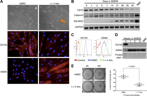
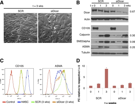
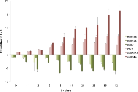
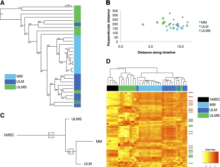
Smooth muscle (SM) is a spontaneously contractile tissue that provides physical support and function to organs such as the uterus. Uterine smooth muscle-related neoplasia comprise common well-differentiated benign lesions called leiomyomas (ULM), and rare, highly aggressive and pleomorphic tumors named leiomyosarcomas (ULMS). MicroRNAs (miRNAs) are small non-coding RNAs that play essential roles in normal cellular development and tissue homeostasis that can be used to accurately subclassify different tumor types. Here, we demonstrate that miRNAs are required for full smooth muscle cell (SMC) differentiation of bone marrow-derived human mesenchymal stem cells (hMSCs). We also report a miRNA signature associated with this process. Moreover, we show that this signature, along with miRNA profiles for ULMS and ULM, are able to subclassify tumors of smooth muscle origin along SM differentiation. Using multiple computational analyses, we determined that ULMS are more similar to hMSCs as opposed to ULM, which are linked with more mature SMCs and myometrium. Furthermore, a comparison of the SM differentiation and ULMS miRNA signatures identified miRNAs strictly associated with SM maturation or transformation, as well as those modulated in both processes indicating a possible dual role. These results support separate origins and/or divergent transformation pathways for ULM and ULMS, resulting in drastically different states of differentiation. In summary, this work expands on our knowledge of the regulation of SM differentiation and sarcoma pathogenesis.


Figures

Figure 1
Figure 1
Cells derived from SM differentiation of hMSCs reproduce the phenotypic and molecular characteristics of mature SMCs. A: Brightfield images (×100 magnification) and immunofluorescence for CD105 and α-smooth muscle actin (ASMA) in human mesenchymal stem cells (hMSC) and after SM differentiation (t = 3 weeks) (×200 magnification). Characteristic ‘hills (orange arrow) and valleys’ are observed by day 21. B: RT-PCR analysis of CD73, calponin, and smooth muscle myosin heavy chain (SM-MHC) during SM differentiation. GAPDH is used as RNA loading control. SMC: RNA from smooth muscle tissue used as a control. FACS (C) and Western blot (D) analyses of CD105 and ASMA during SM differentiation. For FACS, an isotypic control for each respective antibody is used to detect nonspecific staining (Control). SMC: protein lysate of smooth muscle cells isolated from human myometrial tissue. Amido black (protein staining) serves as loading control. E: Gel-contraction assay on hMSCs maintained in MSC medium (hMSC) or smooth muscle differentiation medium (t = 3 weeks), visualized at 0 hours and 12 hours post release (pictures, left); differences in contraction quantified at 12 hours (right). Image J software was used to measure the area of the gel and of the well, which was used for normalization.
Figure 2
Figure 2
miRNAs are required for full SM differentiation. A: Brightfield images of hMSCs transfected with scramble (SCR) (left) or siDicer (right) oligonucleotides (oligo) at t = 3 weeks (×100 magnification) B: Western blot analysis of Dicer, CD105, calponin, SM22α, and ASMA at different time points of SM differentiation, upon Dicer silencing. Actin or tubulin is used as loading control. Numbers on the right indicate the level of reduction of protein levels at t = 3 weeks compared with the corresponding scramble control, after normalization to actin or tubulin. C: FACS analysis of CD105 and ASMA expression in SCR- and siDicer-transfected cells at t = 3 weeks, and hMSC (t = 0) used as control. Control: isotype control. D: qRT-PCR of SM-MHC in hMSCs transfected with scramble oligo or siDicer. Data were normalized to GAPDH expression. Error bars represent standard deviation (SD) between two experimental replicates. FC indicates fold change.
Figure 3
Figure 3
Validation of miRNA array data in SM time course. Fold change (FC) of miRNAs monotonically increasing (red color palette) or decreasing (green palette) during SM differentiation of hMSCs, relative to t = 0, as measured by qRT-PCR. RNU44 was used for normalization. Error bars represent SD between two experimental replicates.
Figure 4
Figure 4
Phylogenetic and clustering analyses of SM tumor subtypes using miRNA profiles. A: Cluster analysis of ULMS, ULM, and MM samples (n = 10 each) based on their miRNA expression profiles. Note that miRNAs are able to accurately segregate benign and malignant uterine mesenchymal lesions. B: Distance analysis of ULMS, ULM, and MM along a timeline based on SM miRNA signature. C: Consensus tree derived from phylogenetic analysis of ULMS, ULM, and MM tissues, rooted by hMSC samples. D: Unsupervised clustering analysis of five hMSC samples along with ULMS, ULM, and MM tissue samples based on the expression of the ‘classifier list’ of miRNAs differentially expressed between the three tissue subtypes. Green and blue hash marks (4A and D) represent a ULMS case that was misclassified and later found to be ULM-like. Arrows indicate SM differentiation signature miRNAs (red: up-regulated miRNAs, green: down-regulated miRNAs).

Comment in

Similar articles

Cited by

References

    1. Hornick JL, Fletcher CD. Criteria for malignancy in nonvisceral smooth muscle tumors. Ann Diagn Pathol. 2003;7:60–66. - PubMed
    1. Skubitz KM, Skubitz AP. Differential gene expression in leiomyosarcoma. Cancer. 2003;98:1029–1038. - PubMed
    1. Halayko AJ, Solway J. Molecular mechanisms of phenotypic plasticity in smooth muscle cells. J Appl Physiol. 2001;90:358–368. - PubMed
    1. Colter DC, Class R, DiGirolamo CM, Prockop DJ. Rapid expansion of recycling stem cells in cultures of plastic-adherent cells from human bone marrow. Proc Natl Acad Sci USA. 2000;97:3213–3218. - PMC - PubMed
    1. Dominici M, Le Blanc K, Mueller I, Slaper-Cortenbach I, Marini F, Krause D, Deans R, Keating A, Prockop D, Horwitz E. Minimal criteria for defining multipotent mesenchymal stromal cells. The International Society for Cellular Therapy position statement, Cytotherapy. 2006;8:315–317. - PubMed

Publication types

MeSH terms