. 2010 Aug;177(2):840-53.
doi: 10.2353/ajpath.2010.090918. Epub 2010 Jun 17.

Long-term expression of tissue-inhibitor of matrix metalloproteinase-1 in the murine central nervous system does not alter the morphological and behavioral phenotype but alleviates the course of experimental allergic encephalomyelitis

Affiliations

Long-term expression of tissue-inhibitor of matrix metalloproteinase-1 in the murine central nervous system does not alter the morphological and behavioral phenotype but alleviates the course of experimental allergic encephalomyelitis

Gioia E M Althoff et al. Am J Pathol. 2010 Aug.

Abstract

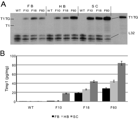
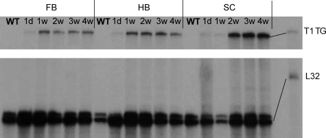
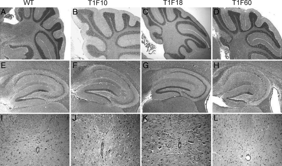
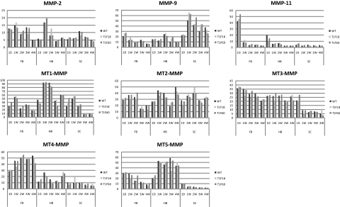
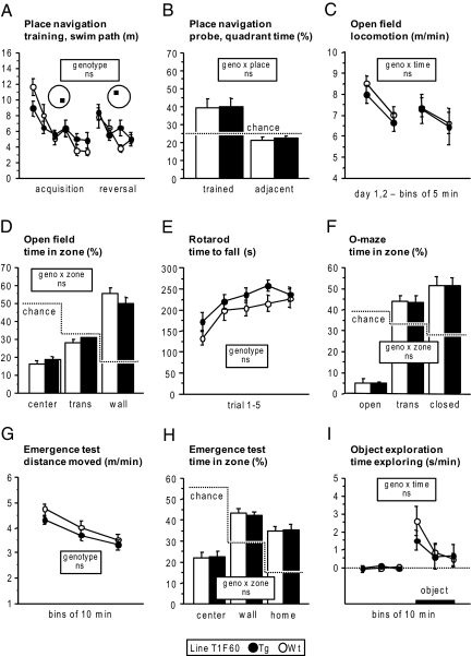
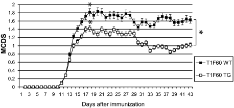
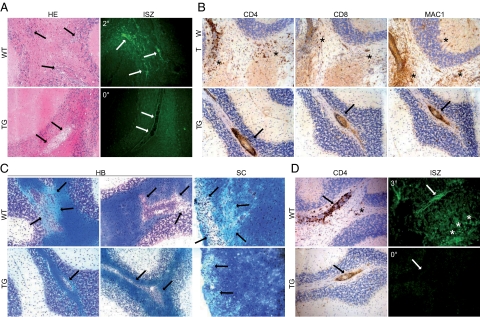
Tissue inhibitors of metalloproteinases (TIMPs) are a family of closely related proteins that inhibit matrix metalloproteinases (MMPs). In the central nervous system (CNS), TIMPs 2, 3, and 4 are constitutively expressed at high levels, whereas TIMP1 can be induced by various stimuli. Here, we studied the effects of constitutive expression of TIMP1 in the CNS in transgenic mice. Transgene expression started prenatally and persisted throughout lifetime at high levels. Since MMP activity has been implicated in CNS development, in proper function of the adult CNS, and in inflammatory disorders, we investigated Timp1-induced CNS alterations. Despite sufficient MMP inhibition, high expressor transgenic mice had a normal phenotype. The absence of compensatory up-regulation of MMP genes in the CNS of Timp1 transgenic mice indicates that development, learning, and memory functions do not require the entire MMP arsenal. To elucidate the effects of strong Timp1 expression in CNS inflammation, we induced experimental allergic encephalomyelitis. We observed a Timp1 dose-dependent mitigation of both experimental allergic encephalomyelitis symptoms and histological lesions in the CNS of transgenic mice. All in all, our data demonstrate that (1) long-term CNS expression of TIMP1 with complete suppression of gelatinolytic activity does not interfere with physiological brain function and (2) TIMP1 might constitute a promising candidate for long-term therapeutic treatment of inflammatory CNS diseases such as multiple sclerosis.


Figures

Figure 1
Figure 1
A: Timp1 mRNA expression in the CNS. An RPA was used to detect Timp1 mRNA in the forebrain (FB), hindbrain (HB), and the spinal cord (SC) of adult animals of the three mouse lines, T1F10, T1F18, and T1F60. Healthy wild-type (WT) animals had no constitutive Timp1 expression. All transgenic mice of the three mouse lines expressed the transgene in every region of the CNS (FB, HB, and SC). The probe consisted of the 3′ portion of Timp1 and an adjacent portion of the hGH polyadenylation signal. This probe allowed for the differentiation of wild-type (T1 264 b) and transgenic Timp1 mRNA (T1 TG 324 b). The RPA was performed as described in Materials and Methods. B: Timp1 concentrations (pg/mg wet tissue) in various CNS regions of transgenic mice of the T1F10, T1F18, and T1F60 lines and WT C57/BL6 mice were determined by enzyme-linked immunosorbent assay. Error bars = SEM.
Figure 2
Figure 2
Postnatal time course of transgenic Timp1 expression in the T1F60 mouse line. Transgene expression (T1 TG) was detectable in every region of the CNS (forebrain [FB], hindbrain [HB], and spinal cord [SC]) on the first postnatal day and reached maximum levels within the first postnatal week. There was no gene expression of wild-type Timp1. The L32 band served as a loading control. RPA was performed as described in Materials and Methods.
Figure 3
Figure 3
In situ hybridization for Timp1. No Timp1 RNA was detected in the brains of adult wild-type animals (AC), whereas strong Timp1 signals were observed in all CNS regions of adult TG T1F60 mice (DF, arrows). Hippocampus (A, D), cerebellum (B, E), and spinal cord (C, F). The Timp1 RNA (arrows) co-localized with GFAP immunoreactive cells (brown staining) demonstrating that transgenic Timp1 was synthesized by astrocytes (G, H). A minority of astrocytes in the T1F60 mouse line were Timp1 RNA negative (G, arrowheads). At embryonic stage E 17.5, few cells showed transgene expression in transgenic mice of the T1F60 mouse line (I, arrows). Wild-type mice of this stage revealed no positive staining in the CNS (data not shown). Original magnification: ×40 (AF); ×100 (GI). In situ zymography on brain sections of P10 mice showed gelatinolytic activity in the forebrain (J), the hindbrain (K), and the spinal cord (L) of wild-type animals (JL, arrows). No activity was detected in the same regions of transgenic littermates of the T1F60 mouse line (MO). Control slices showed that the enzymatic activity was inhibited by 10 mmol/L EDTA and that substrate was necessary to obtain a fluorescent signal (data not shown). Original magnification: ×20.
Figure 4
Figure 4
Histology of the CNS. The morphology of the cerebellum (AD), hippocampus (EH), and spinal cord (IL) of adult animals of all three mouse lines revealed no alterations as compared with WT mice. H&E magnification: ×5 (AH); ×20 (IL).
Figure 5
Figure 5
Postnatal MMP RNA expression in the CNS. RPA was performed on CNS tissue from forebrain (FB), hindbrain (HB), and spinal cord (SC) of 1 day (1D), 1-, 2-, 3-, and 4-week-old (1W, 2W, 3W, and 4W, respectively) animals. The amount of specific RNA is depicted as arbitrary units (y axis). Patterns of regulation are represented for MMP-2, MMP-9, MMP-11, and MT1-MMPs to MT5-MMPs. RPA was performed as described in Materials and Methods.
Figure 6
Figure 6
Behavioral tests. A and B: Water-maze place navigation: A: Overall performance and learning rates were similar in wild-type (WT) and TG mice of line T1F60. Each point represents three subsequent trials: genotype F(1,25) = 0.025 ns; time F(9225) = 8.775, P < 0.0001; genotype × time F(9225) = 1.598 ns. B: Irrespective of genotype, mice of line T1F60 showed a robust preference for the target quadrant in comparison with the adjacent quadrants: place F(1,25) = 16.108, P < 0.0005; genotype × place F(1,25) = 0.013 ns. CI: Anxiety-related behaviors, exploratory activity, and locomotion: C: Wild-type and TG of line T1F60 did not differ with respect to level and time course of activity: genotype F(1,25) = 0.271 ns; time F(3,75) = 9.293, P < 0.0001; genotype × time F(3,75) = 0.150 ns. D: Time spent in the center, transition, and wall zone of the open field. Independently of genotype, mice avoided the center zone in favor of the wall zone: zone F(2,52) = 68.500, P < 0.0001; genotype × zone F(2,52) = 1.198 ns. E: TG and wild-type were indistinguishable with respect to both performance level and learning rate in the rotarod test: genotype F(1,26) = 2.162 ns; trial F(4104) = 11.875, P < 0.0001; genotype × trial F(4104) = 0.492 ns. F: Time spent in the open, transition, and closed zone of the elevated O-maze. Independently of genotype, mice strongly avoided the open zone: zone F(2,52) = 89.770, P < 0.0001; genotype × zone F(2,52) = 0.007 ns. G: Although TG moved on average slightly shorter distances than wild-type, there was no genotype effect on level or habituation rate of activity: genotype F(1,26) = 1.692 ns; time F(2,52) = 47.435, P < 0.0001; genotype × time F(2,52) = 0.570 ns. H: Irrespective of genotype, mice preferred the home and wall zones: zone F(2,52) = 27.386, P < 0.0001; genotype × zone F(2,52) = 0.089 ns. I: Exploratory activity toward a novel object was also significant and unaffected by genotype: genotype F(1,26) = 0.233 ns; time F(5130) = 8.079, P < 0.0001; genotype × time F(5130) = 0.668 ns.
Figure 7
Figure 7
Disease course in the EAE model of wild-type (WT) and transgenic mice of the T1F60 (nWT = 31; nTG = 31) line. 0: no symptoms; 1: limp tail; 2: ataxia; 3: hind limb paresis; 4: hind limb and fore limb paresis; 5: moribund; *P ≤ 0.05.
Figure 8
Figure 8
Pattern of leukocyte infiltration, gelatinolytic activity, and demyelination in the hindbrain of wild-type (WT) and transgenic (TG) mice of the T1F60 mouse line after induction of EAE. At day 15 after immunization wild-type animals with a clinical score of 2 (2°) showed diffuse lymphocytic infiltration (A, HE arrows) correlating with strong gelatinolytic activity detected by in situ zymography (ISZ) in serial sections (A, ISZ arrows). Despite a clinical score of 0 (0°), TG animals revealed perivascular accumulation of lymphocytes (A, HE arrows) and no gelatinolytic activity (A, ISZ arrows). At 40 days there was diffuse infiltration of CD4+ and CD8+ lymphocytes as well as MAC1+ macrophages and reactive microglia in symptomatic control animals (B, wild-type, asterisks). In transgenic mice these cells were predominantly confined to perivascular cuffs (B, TG, arrows). Klüver-Barrera (KB) staining revealed demyelinated areas in the white matter of hindbrain (HB) and spinal cord (SC) of wild-type mice (C, wild-type, arrows). In transgenic T1F60 mice there was an overall normal myelinization around inflammatory lesions in the cerebellum and spinal cord (C, TG, arrow). At day 40 wild-type animals with a high clinical disease score of 3 (3°) showed strong perivascular and diffuse infiltration of CD4+ lymphocytes (D, wild-type CD4, arrow and asterisk, respectively) correlating to perivascular and diffuse gelatinolytic activity (D, wild-type ISZ arrow and asterisks, respectively). In sharp contrast, CD4+ lymphocytes accumulated around the vessels in TG hindbrain and no gelatinolysis was observed (D, TG CD4 and ISZ arrows). Addition of 10 mmol/L EDTA or omission of the quenched gelatin resulted in no fluorescent signal (not shown). Original magnification: ×40 (A and B); ×20 (C and D).

Similar articles

Cited by

References

    1. Edwards D, Waterhouse P, Holman M, Denhardt D. A growth-responsive gene (16C8) in normal mouse fibroblasts homologous to a human collagenase inhibitor with erythroid-potentiating activity: evidence for inducible and constitutive transcripts. Nucleic Acids Res. 1986;14:8863–8878. - PMC - PubMed
    1. Leco K, Hayden L, Sharma R, Rocheleau H, Greenberg A, Edwards D. Differential regulation of TIMP-1 and TIMP-2 mRNA expression in normal and Ha-ras-transformed murine fibroblasts. Gene. 1992;117:209–217. - PubMed
    1. Sternlicht M, Werb Z. How matrix metalloproteinases regulate cell behavior. Annu Rev Cell Dev Biol. 2001;17:463–516. - PMC - PubMed
    1. Vaillant C, Didier-Bazès M, Hutter A, Belin M, Thomasset N. Spatiotemporal expression patterns of metalloproteinases and their inhibitors in the postnatal developing rat cerebellum. J Neurosci. 1999;19:4994–5004. - PMC - PubMed
    1. Ulrich R, Gerhauser I, Seeliger F, Baumgärtner W, Alldinger S. Matrix metalloproteinases and their inhibitors in the developing mouse brain and spinal cord: a reverse transcription quantitative polymerase chain reaction study. Dev Neurosci. 2005;27:408–418. - PubMed

Publication types

MeSH terms

Substances