

A septin diffusion barrier at the base of the primary cilium maintains ciliary membrane protein distribution
- PMID: 20558667
- PMCID: PMC3092790
- DOI: 10.1126/science.1191054
A septin diffusion barrier at the base of the primary cilium maintains ciliary membrane protein distribution
Abstract
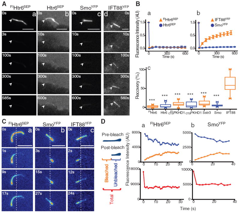
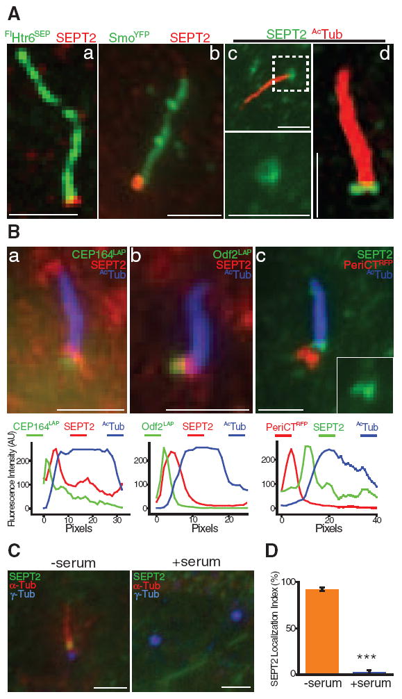
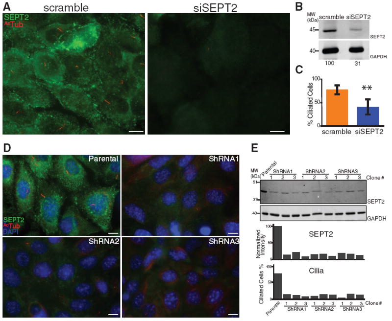
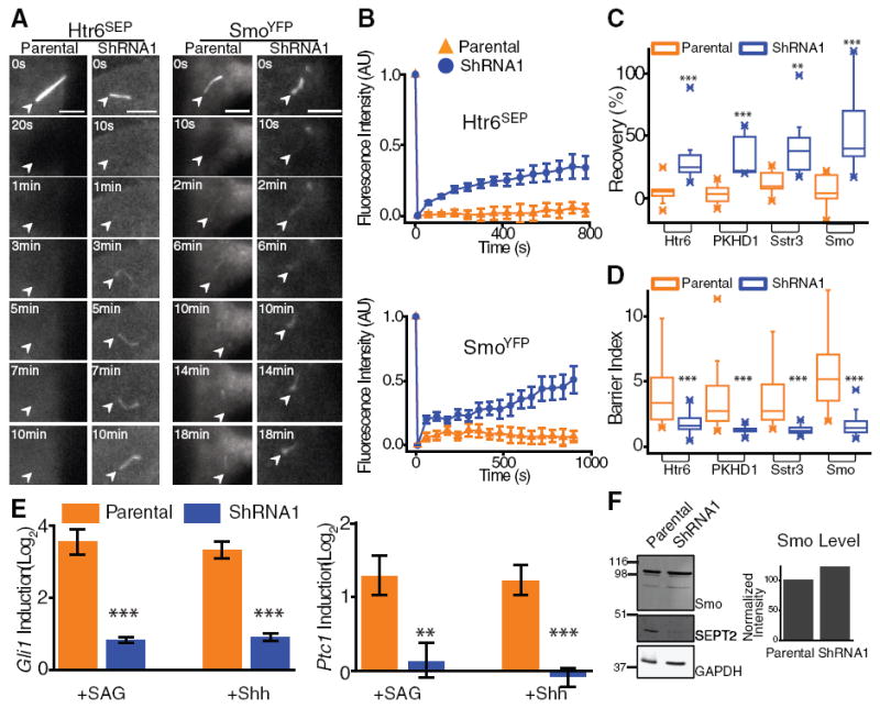
In animal cells, the primary cilium transduces extracellular signals through signaling receptors localized in the ciliary membrane, but how these ciliary membrane proteins are retained in the cilium is unknown. We found that ciliary membrane proteins were highly mobile, but their diffusion was impeded at the base of the cilium by a diffusion barrier. Septin 2 (SEPT2), a member of the septin family of guanosine triphosphatases that form a diffusion barrier in budding yeast, localized at the base of the ciliary membrane. SEPT2 depletion resulted in loss of ciliary membrane protein localization and Sonic hedgehog signal transduction, and inhibited ciliogenesis. Thus, SEPT2 is part of a diffusion barrier at the base of the ciliary membrane and is essential for retaining receptor-signaling pathways in the primary cilium.
Figures
Comment in
-
Cytoskeleton: Breaking the diffusion barrier.Nat Rev Mol Cell Biol. 2010 Aug;11(8):542. doi: 10.1038/nrm2945. Nat Rev Mol Cell Biol. 2010. PMID: 20651705 No abstract available.
-
Cell biology. Septins at the nexus.Science. 2010 Sep 10;329(5997):1289-90. doi: 10.1126/science.1195445. Science. 2010. PMID: 20829470 No abstract available.
Similar articles
-
Ciliary diffusion barrier: the gatekeeper for the primary cilium compartment.Cytoskeleton (Hoboken). 2011 Jun;68(6):313-24. doi: 10.1002/cm.20514. Epub 2011 Jun 10. Cytoskeleton (Hoboken). 2011. PMID: 21634025 Free PMC article. Review.
-
Septins 2, 7 and 9 and MAP4 colocalize along the axoneme in the primary cilium and control ciliary length.J Cell Sci. 2013 Jun 15;126(Pt 12):2583-94. doi: 10.1242/jcs.111377. Epub 2013 Apr 9. J Cell Sci. 2013. PMID: 23572511 Free PMC article.
-
The role of ciliary trafficking in Hedgehog receptor signaling.Sci Signal. 2015 Jun 2;8(379):ra55. doi: 10.1126/scisignal.aaa5622. Sci Signal. 2015. PMID: 26038600 Free PMC article.
-
Patched1 regulates hedgehog signaling at the primary cilium.Science. 2007 Jul 20;317(5836):372-6. doi: 10.1126/science.1139740. Science. 2007. PMID: 17641202
-
The base of the cilium: roles for transition fibres and the transition zone in ciliary formation, maintenance and compartmentalization.EMBO Rep. 2012 Jun 29;13(7):608-18. doi: 10.1038/embor.2012.73. EMBO Rep. 2012. PMID: 22653444 Free PMC article. Review.
Cited by
-
Keeping the balance between proliferation and differentiation: the primary cilium.Curr Genomics. 2011 Jun;12(4):285-97. doi: 10.2174/138920211795860134. Curr Genomics. 2011. PMID: 22131874 Free PMC article.
-
Protein sorting, targeting and trafficking in photoreceptor cells.Prog Retin Eye Res. 2013 Sep;36:24-51. doi: 10.1016/j.preteyeres.2013.03.002. Epub 2013 Apr 3. Prog Retin Eye Res. 2013. PMID: 23562855 Free PMC article. Review.
-
Septin Form and Function at the Cell Cortex.J Biol Chem. 2015 Jul 10;290(28):17173-80. doi: 10.1074/jbc.R114.634444. Epub 2015 May 8. J Biol Chem. 2015. PMID: 25957401 Free PMC article. Review.
-
Regulated membrane protein entry into flagella is facilitated by cytoplasmic microtubules and does not require IFT.Curr Biol. 2013 Aug 5;23(15):1460-5. doi: 10.1016/j.cub.2013.06.025. Epub 2013 Jul 25. Curr Biol. 2013. PMID: 23891117 Free PMC article.
-
Steric volume exclusion sets soluble protein concentrations in photoreceptor sensory cilia.Proc Natl Acad Sci U S A. 2012 Jan 3;109(1):203-8. doi: 10.1073/pnas.1115109109. Epub 2011 Dec 19. Proc Natl Acad Sci U S A. 2012. PMID: 22184246 Free PMC article.
References
-
- Marshall WF, Nonaka S. Curr Biol. 2006;16:R604. - PubMed
Publication types
MeSH terms
Substances
Grants and funding
LinkOut - more resources
Full Text Sources
Other Literature Sources
Molecular Biology Databases
