. 2010 Sep 3;285(36):27553-60.
doi: 10.1074/jbc.M110.144394. Epub 2010 Jun 17.

Francisella tularensis antioxidants harness reactive oxygen species to restrict macrophage signaling and cytokine production

Affiliations

Francisella tularensis antioxidants harness reactive oxygen species to restrict macrophage signaling and cytokine production

Amanda A Melillo et al. J Biol Chem. .

Abstract

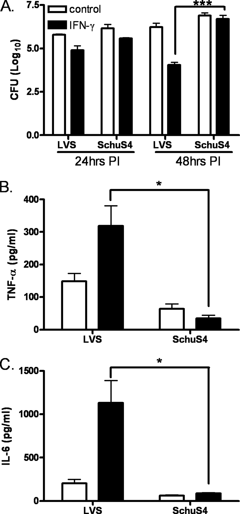
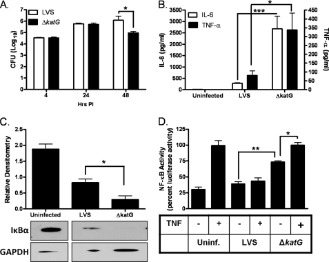
Francisella tularensis is the etiologic agent of the highly infectious animal and human disease tularemia. Its extreme infectivity and virulence are associated with its ability to evade immune detection, which we now link to its robust reactive oxygen species-scavenging capacity. Infection of primary human monocyte-derived macrophages with virulent F. tularensis SchuS4 prevented proinflammatory cytokine production in the presence or absence of IFN-gamma compared with infection with the attenuated live vaccine strain. SchuS4 infection also blocked signals required for macrophage cytokine production, including Akt phosphorylation, IkappaB alpha degradation, and NF-kappaB nuclear localization and activation. Concomitant with SchuS4-mediated suppression of Akt phosphorylation was an increase in the levels of the Akt antagonist PTEN. Moreover, SchuS4 prevented the H(2)O(2)-dependent oxidative inactivation of PTEN compared with a virulent live vaccine strain. Mutation of catalase (katG) sensitized F. tularensis to H(2)O(2) and enhanced PTEN oxidation, Akt phosphorylation, NF-kappaB activation, and inflammatory cytokine production. Together, these findings suggest a novel role for bacterial antioxidants in restricting macrophage activation through their ability to preserve phosphatases that temper kinase signaling and proinflammatory cytokine production.


Figures

FIGURE 1.
FIGURE 1.
SchuS4 inhibits proinflammatory cytokines in unstimulated and IFN-γ-stimulated macrophages. hMDMs were infected with LVS or SchuS4. Cells were lysed at 24 or 48 h post-infection (PI), and intracellular bacteria were quantified (A). TNF-α (B) and IL-6 (C) levels were measured in the culture supernatants 24 h post-infection using the BDTM cytometric bead array. The statistical significance of the results was examined with Student's t test, and p values were recorded. *, p < 0.05; ***, p < 0.001 compared with infection with LVS (n = 6). CFU, colony-forming units.
FIGURE 2.
FIGURE 2.
SchuS4 restricts NF-κB and Akt signaling pathways. A, hMDMs were stimulated with IFN-γ for 18 h prior to infection with LVS or SchuS4 for the indicated times. IκBα degradation and phosphorylation were determined by Western blot analysis. U, uninfected; GAPDH, glyceraldehyde-3-phosphate dehydrogenase. B, shown are the results from the densitometric analysis of IκBα degradation 30 min post-infection (n = 3). C, shown is the nuclear translocation of the NF-κB p65 subunit in IFN-γ-simulated hMDMs infected with LVS or SchuS4 with or without 10 ng of TNF-α for 20 min post-infection by immunofluorescence. D, the quantification of p65 localization was calculated and is expressed as the ratio of nuclear to whole cell staining (n = 20). E, IFN-γ-stimulated hMDMs were infected with LVS or SchuS4 for 1 or 4 h. Erk phosphorylation and LKB-1 levels were assayed by Western blotting. hMDMs were infected with LVS or SchuS4 for 1 h. Total and phospho-PTEN and total and phospho-Akt levels were monitored by Western blot analysis. hpi, hours post-infection. G, shown are the results from densitometric analysis of the bands shown in F expressed as ratios of total PTEN to phospho-PTEN and of phospho-Akt to total Akt. The data are cumulative of three independent experiments. The statistical analyses were performed by the Tukey-Kramer multiple comparison test, and p values were recorded. **, p < 0.01; ***, p < 0.001.
FIGURE 3.
FIGURE 3.
F. tularensis modulates PTEN oxidation and Akt phosphorylation. A, A600 of F. tularensis LVS and SchuS4 following 18 h of treatment with the indicated concentrations of H2O2. Data represent the mean ± S.E. of quadruplicate samples. PI, post-infection. B, infection of HT1080 cells with LVS, ΔkatG, or SchuS4 for 1 h. 300 μm H2O2 was added (3 mm H2O2 was added in the lower panel). PTEN oxidation and Akt phosphorylation were determined by Western blot analysis. Uninf., uninfected. C, densitometric analysis of reduced (re) and oxidized (ox) PTEN expressed as percent of total PTEN (n = 3). D, Western blot analysis of PTEN oxidation and Akt phosphorylation in hMDMs infected with LVS (L), ΔkatG (K), or SchuS4 (S) for 1 h and treated with 300 μm H2O2. Statistical analyses were performed by the Tukey-Kramer multiple comparison test, and p values were recorded. *, p < 0.05. U, uninfected; GAPDH, glyceraldehyde-3-phosphate dehydrogenase.
FIGURE 4.
FIGURE 4.
F. tularensis catalase protects from H2O2 toxicity and restricts macrophage H2O2 production. A, shown are A600 measurements of F. tularensis LVS and ΔkatG following 22 h of treatment with the indicated concentrations of H2O2. PI, post-infection. B, intracellular H2O2 levels were calculated as described under “Experimental Procedures.” Data points were normalized by dividing with catalase (Cat) activity measured at time 0. The data represent the mean ± S.E. of three independent experiments. C, U937 cells transfected with roGFP1 were infected with LVS or ΔkatG. GFP fluorescence resulting from excitation at either 400 or 470 nm was monitored at the indicated times. The results are expressed as relative to the ratios of the 400/470 nm spectra. Increases in the 400/470 nm ratio are indicative of greater oxidation. The statistical significance of the results was examined by Student's t test, and p values were recorded. *, p < 0.05; **, p < 0.01 compared with infection with LVS. Ox, oxidized; Red, reduced.
FIGURE 5.
FIGURE 5.
Loss of F. tularensis catalase impairs the cytokine-suppressing capacity of F. tularensis. Unstimulated hMDMs were infected with LVS or ΔkatG. A, the cells were lysed at 24 h post-infection (PI), and intracellular bacteria were quantified. CFU, colony-forming units. B, IL-6 and TNF-α levels were measured in cell culture supernatants (n = 6) 24 h post-infection using the BDTM cytometric bead array. C, hMDMs were infected with LVS or ΔkatG for 20 min. IκBα degradation was determined by Western blot analysis. GAPDH, glyceraldehyde-3-phosphate dehydrogenase. D, shown is the luciferase activity in HT1080 cells transfected with the NF-κB-luciferase construct and infected with LVS or ΔkatG for 4 h. TNF-α (10 ng) was added after 2 h of infection where indicated. The data are expressed as percent luciferase activity and represent the mean ± S.E. from three independent experiments. The statistical significance of the results was examined with the Student's t test, and p values were recorded. *, p < 0.05; **, p < 0.01; ***, p < 0.001 compared with infection with LVS. Uninf., uninfected.

Similar articles

Cited by

References

    1. Oyston P. C., Sjostedt A., Titball R. W. (2004) Nat. Rev. Microbiol. 2, 967–978 - PubMed
    1. Telepnev M., Golovliov I., Sjöstedt A. (2005) Microb. Pathog. 38, 239–247 - PubMed
    1. Lauriano C. M., Barker J. R., Yoon S. S., Nano F. E., Arulanandam B. P., Hassett D. J., Klose K. E. (2004) Proc. Natl. Acad. Sci. U.S.A. 101, 4246–4249 - PMC - PubMed
    1. Rajaram M. V., Ganesan L. P., Parsa K. V., Butchar J. P., Gunn J. S., Tridandapani S. (2006) J. Immunol. 177, 6317–6324 - PubMed
    1. Parsa K. V., Ganesan L. P., Rajaram M. V., Gavrilin M. A., Balagopal A., Mohapatra N. P., Wewers M. D., Schlesinger L. S., Gunn J. S., Tridandapani S. (2006) PLoS Pathog. 2, e71. - PMC - PubMed

Publication types

MeSH terms