. 2010 Aug 13;285(33):25831-40.
doi: 10.1074/jbc.M110.112128. Epub 2010 Jun 17.

BCL-3 degradation involves its polyubiquitination through a FBW7-independent pathway and its binding to the proteasome subunit PSMB1

Affiliations

BCL-3 degradation involves its polyubiquitination through a FBW7-independent pathway and its binding to the proteasome subunit PSMB1

Aurore Keutgens et al. J Biol Chem. .

Abstract

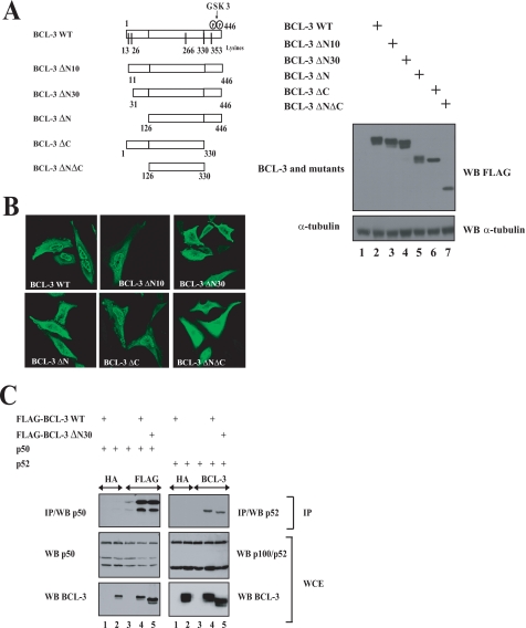
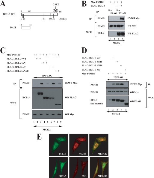
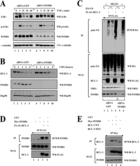
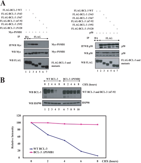
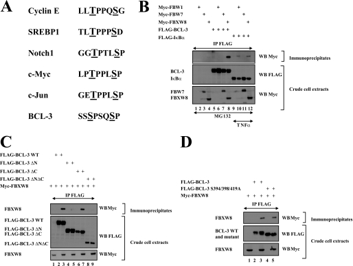
The oncogenic protein BCL-3 activates or represses gene transcription through binding with the NF-kappaB proteins p50 and p52 and is degraded through a phospho- and GSK3-dependent pathway. However, the mechanisms underlying its degradation remain poorly understood. Yeast two-hybrid analysis led to the identification of the proteasome subunit PSMB1 as a BCL-3-associated protein. The binding of BCL-3 to PSMB1 is required for its degradation through the proteasome. Indeed, PSMB1-depleted cells are defective in degrading polyubiquitinated BCL-3. The N-terminal part of BCL-3 includes lysines 13 and 26 required for the Lys(48)-linked polyubiquitination of BCL-3. Moreover, the E3 ligase FBW7, known to polyubiquitinate a variety of substrates phosphorylated by GSK3, is dispensable for BCL-3 degradation. Thus, our data defined a unique motif of BCL-3 that is needed for its recruitment to the proteasome and identified PSMB1 as a key protein required for the proteasome-mediated degradation of a nuclear and oncogenic IkappaB protein.


Figures

FIGURE 1.
FIGURE 1.
The N-terminal domain of BCL-3 harbors a nuclear localization signal. A, left, schematic representation of the BCL-3 constructs used in immunofluorescence analysis. The GSK3 phosphorylation sites (Ser394 and Ser398) as well as the lysine residues (13, 26, 266, 330, and 353) on BCL-3 are illustrated. On the right, anti-FLAG and α-tubulin (loading control) Western blots using extracts from 293 cells transfected with the indicated expression plasmids. B, the N-terminal domain of BCL-3 is critical for its nuclear localization. HeLa cells were transfected with the indicated FLAG-tagged expression plasmids, and immunofluorescence studies were carried out using the anti-FLAG antibody. C, the 30 N-terminal amino acids of BCL-3 are dispensable for the interaction with p50 and p52. 293 cells were transfected with the indicated expression plasmids, and cell extracts were subjected to anti-HA (negative control), anti-FLAG, or anti-BCL-3 immunoprecipitations (IP) followed by anti-p50 or anti-p100/p52 Western blots (WB; left and right top panels, respectively). Anti-p100/p52, anti-p50, and anti-BCL-3 Western blots were carried out with crude cell extracts (WCE) as well (bottom panels).
FIGURE 2.
FIGURE 2.
BCL-3 binding to p50 or p52 is required for constitutive BCL-3 phosphorylation. A, mutation of residues within the second ankyrin repeats of BCL-3. Both IκBα and BCL-3 are illustrated schematically. The NLS sequence within the second ankyrin repeat of IκBα is shown. The FLAG-IκBα NLS MT construct encodes a FLAG-tagged IκBα protein harboring three mutations within the NLS sequence (23). Single, double, or triple mutants where the corresponding leucine and isoleucine residues within the second ankyrin repeats of BCL-3 were mutated into alanines, as indicated (BCL-3 ANK M1, -M12, or M123, respectively). B and C, the integrity of the second ankyrin repeat of BCL-3 is required for binding to p50 (B) or p52 (C). 293 cells were transfected with the indicated expression plasmids and anti-HA (negative control) -FLAG, or -BCL-3 immunoprecipitations (IP) followed by anti-p50 or p52 Western blot analysis were carried out (top panels). Cell extracts also were subjected to anti-FLAG, -p50, and -p52 Western blot analysis (bottom panels). D, mutations within the second ankyrin repeat alter the nuclear localization of BCL-3. HeLa cells were transfected with the indicated expression plasmids and the resulting cells were subjected to immunofluorescence analysis using the anti-FLAG antibody. E, constitutive GSK3-mediated BCL-3 phosphorylation in the cytoplasm and in the nucleus of Karpas cells. Lymphoma-derived Karpas cells were left untreated (lanes 1, 2, 5, and 6) or stimulated with LiCl for the indicated periods of times (lanes 3, 4, 7, and 8), and cell extracts were subjected to anti-HA (negative control, lanes 1 and 5) or anti-BCL-3 immunoprecipitations (lanes 2–4 and 6–8) followed by anti-p52/p100 Western blot (WB) analysis (top panel). Crude cell extracts were subjected to anti-p52/p100, anti-BCL-3, α-tubulin and anti-NBS1 Western blots as well (bottom panels). WCE, whole cell extract.
FIGURE 3.
FIGURE 3.
The degradation of BCL-3 through the proteasome requires lysines 13 and 26. A and B, the proteasome is required for BCL-3 degradation. 293 cells were transfected with the indicated expression plasmids and subsequently left untreated or stimulated for 2 h with MG132 (20 μm) the next day. Cell extracts (non-SDS lysis buffer (lanes 1–4 (A)) or SDS lysis buffer (A, lanes 5–6, and B)) were subjected to anti-FLAG immunoprecipitations (IP) followed by anti-HA (A and B) and anti-p50 (A) Western blot (WB) analysis (top panels). Cell extracts also were subjected to anti-FLAG (A and B), anti-p50 (A), and anti-HA (A and B) Western blot analysis (bottom panels). C, critical roles of lysines 13 and 26 for the Lys48-linked polyubiquitination (poly-Ub) of BCL-3. 293 cells were transfected with the indicated expression plasmids and subsequently treated with MG132 (20 μm) for 4 h. Cell extracts were subjected to anti-BCL-3 immunoprecipitations followed by anti-Myc Western blot analysis (top panel). Anti-BCL-3 and anti-Myc Western blots also were carried out on the crude cell extracts (bottom panels). D, subcellular localization of wild type and BCL-3 mutants. HeLa cells were transfected with the indicated FLAG-tagged expression plasmid, and immunofluorescence studies were carried out using the anti-FLAG antibody. WCE, whole cell extract.
FIGURE 4.
FIGURE 4.
PSMB1 is a BCL-3-interacting protein. A, a schematic representation of both BCL-3 and the bait used for yeast two-hybrid analyses. The GSK3 phosphorylation sites (Ser394 and Ser398) as well as the lysine residues (13, 26, 266, 330, and 353) on BCL-3 are illustrated. B–D, ectopically expressed BCL-3 and PSMB1 interact in mammalian cells through the N-terminal domain of BCL-3. 293 cells were transfected with the indicated expression plasmids. Cells were treated with MG132 (20 μm) for 4 h, and anti-HA (B) (negative control) or anti-FLAG (B–D) immunoprecipitations (IP) followed by an anti-Myc Western blot (WB) performed on the immunoprecipitates were carried out (top panels). Crude cell extracts were subjected to anti-Myc and anti-FLAG Western blots as well. E, PMSB1 and BCL-3 mainly co-localize in the nucleus. HeLa cells were transfected with FLAG-BCL-3 and with Myc-PMSB1, and their localizations were revealed through anti-FLAG and anti-Myc immunofluorescence, respectively. PML bodies also were visualized using the corresponding anti-PML antibody. WCE, whole cell extract.
FIGURE 5.
FIGURE 5.
PSMB1-deficient cells show impaired BCL-3 degradation. A, PSMB1 depletion interferes with TNFα-mediated IκBα degradation but not its phosphorylation. 293 cells were transfected with a siRNA targeting either GFP (negative control, lanes 1–5) or PSMB1 (lanes 6–10). Cells subsequently were left untreated (lanes 1 and 6) or stimulated with TNFα (100 units/ml) for the indicated periods of time. Cell extracts were subjected to anti-IκBα, -phospho-IκBα, -PSMB1, and α-tubulin, as indicated. B, BCL-3 half-life is increased upon PSMB1 depletion. Karpas cells were transfected with the indicated siRNA and subsequently left untreated (lanes 1 and 6) or stimulated with cycloheximide (CHX; 50 μg/ml) (lanes 2–5 and 7–10) for the indicated periods of time. Anti-BCL-3, -PSMB1, and -Hsp90 (used for normalization purposes) Western blots (WB) were carried out on the crude cell extracts, as indicated. C, Lys48-linked polyubiquitinated (poly-Ub) forms of BCL-3 are accumulating upon MG132 treatment or PSMB1 depletion. siRNA GFP or PSMB1 293 cells were transfected with the indicated expression plasmids and subsequently left untreated (lanes 1, 2, 5, and 6) or stimulated with MG132 (20 μm) (lanes 3 and 4) for 4 h. Cell extracts were subjected to anti-FLAG immunoprecipitates (IP) followed by anti-HA Western blot analysis (top panel). Anti-HA, -FLAG, -NBS1, and -PSMB1 Western blots also were carried out with the crude cell extracts (bottom panels). D and E, GSK3 enhances the association of BCL-3 to the proteasome. 293 cells were transfected with the indicated expression plasmids and subsequently left untreated (lanes 1 and 3 (D) and lanes 1, 2, and 4 (E)) or stimulated with LiCl (50 mm) for 2 h (lanes 2 and 4 (D) and lane 3 (E)). Anti-FLAG (D) or -Myc (E) immunoprecipitates were subjected to anti-PSMB1 (D) or BCL-3 (E) Western blot analysis (top panels). Crude cell extracts also were subjected to anti-PSMB1, -FLAG (D), and -BCL-3 (E) Western blots (bottom panels). WCE, whole cell extract.
FIGURE 6.
FIGURE 6.
PSMB1 is required for BCL-3 degradation. A, identification of the PSMB1-binding domain on BCL-3. 293 cells were transfected with the indicated expression plasmids an anti-HA (negative control) or -FLAG immunoprecipitations (IP), followed by anti-Myc or -p50 Western blots (WB), were carried out (left top panels and right top panels, respectively). Crude cell extracts were subjected to anti-Myc, -FLAG, and -p50 Western blots, as indicated (bottom panels). B, increased half-life of the BCL-3 mutant that fails to bind PSMB1 (BCL-3 Δ67–92 also named BCL-3 ΔPSMB1). WT or the BCL-3 ΔPSMB1 mutant were transfected in 293 cells, and the resulting cells were left untreated or stimulated with cycloheximide (CHX) for the indicated periods of time. Anti-BCL-3 and -Hsp90 (loading control) Western blots were performed on the crude cell extracts. Bottom panel, a quantification of WT BCL-3 or BCL-3 ΔPSMB1 levels is shown in transfected 293 cells. The signal intensity in unstimulated 293 cells is set to 100% for both experimental conditions.
FIGURE 7.
FIGURE 7.
FBW1, FBW7, and FBXW8 are dispensable for BCL-3 degradation through the GSK3-dependent pathway. A, listing of the phosphodegrons seen in multiple FBW7 substrates and in BCL-3. The residues phosphorylated by GSK3 are underlined. B–D, FBXW8 but not FBW1 and FBW7 bind BCL-3. 293 cells were transfected with the indicated expression constructs and subsequently left untreated (C and D) or stimulated with MG132 and/or TNFα as indicated (B). Anti-FLAG immunoprecipitations followed by anti-Myc Western blot analysis were performed (top panels). Crude cell extracts were subjected to anti-FLAG and -Myc Western blots as well (bottom panels).

Similar articles

Cited by

References

    1. Bours V., Franzoso G., Azarenko V., Park S., Kanno T., Brown K., Siebenlist U. (1993) Cell 72, 729–739 - PubMed
    1. Palmer S., Chen Y. H. (2008) Immunol. Res. 42, 210–218 - PubMed
    1. Franzoso G., Bours V., Azarenko V., Park S., Tomita-Yamaguchi M., Kanno T., Brown K., Siebenlist U. (1993) EMBO J. 12, 3893–3901 - PMC - PubMed
    1. Kerr L. D., Duckett C. S., Wamsley P., Zhang Q., Chiao P., Nabel G., McKeithan T. W., Baeuerle P. A., Verma I. M. (1992) Genes Dev. 6(12A), 2352–2363 - PubMed
    1. Nolan G. P., Fujita T., Bhatia K., Huppi C., Liou H. C., Scott M. L., Baltimore D. (1993) Mol. Cell. Biol. 13, 3557–3566 - PMC - PubMed

Publication types

MeSH terms