

Biophysical investigations of complement receptor 2 (CD21 and CR2)-ligand interactions reveal amino acid contacts unique to each receptor-ligand pair
- PMID: 20558730
- PMCID: PMC2930724
- DOI: 10.1074/jbc.M110.106617
Biophysical investigations of complement receptor 2 (CD21 and CR2)-ligand interactions reveal amino acid contacts unique to each receptor-ligand pair
Abstract
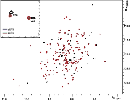
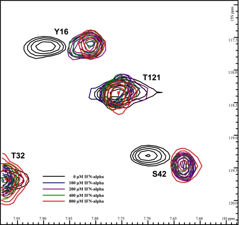
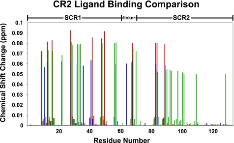
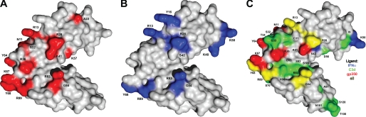
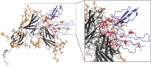
Human complement receptor type 2 (CR2 and CD21) is a cell membrane receptor, with 15 or 16 extracellular short consensus repeats (SCRs), that promotes B lymphocyte responses and bridges innate and acquired immunity. The most distally located SCRs, SCR1-2, mediate the interaction of CR2 with its four known ligands (C3d, EBV gp350, IFNalpha, and CD23). To ascertain specific interacting residues on CR2, we utilized NMR studies wherein gp350 and IFNalpha were titrated into (15)N-labeled SCR1-2, and chemical shift changes indicative of specific inter-molecular interactions were identified. With backbone assignments made, the chemical shift changes were mapped onto the crystal structure of SCR1-2. With regard to gp350, the binding region of CR2 is primarily focused on SCR1 and the inter-SCR linker, specifically residues Asn(11), Arg(13), Ala(22), Arg(28), Ser(32), Arg(36), Lys(41), Lys(57), Tyr(64), Lys(67), Tyr(68), Arg(83), Gly(84), and Arg(89). With regard to IFNalpha, the binding is similar to the CR2-C3d interaction with specific residues being Arg(13), Tyr(16), Arg(28), Ser(42), Lys(48), Lys(50), Tyr(68), Arg(83), Gly(84), and Arg(89). We also report thermodynamic properties of each ligand-receptor pair determined using isothermal titration calorimetry. The CR2-C3d interaction was characterized as a two-mode binding interaction with K(d) values of 0.13 and 160 microm, whereas the CR2-gp350 and CR2-IFNalpha interactions were characterized as single site binding events with affinities of 0.014 and 0.035 microm, respectively. The compilation of chemical binding maps suggests specific residues on CR2 that are uniquely important in each of these three binding interactions.
Figures
Similar articles
-
Mapping of the C3d ligand binding site on complement receptor 2 (CR2/CD21) using nuclear magnetic resonance and chemical shift analysis.J Biol Chem. 2009 Apr 3;284(14):9513-20. doi: 10.1074/jbc.M808404200. Epub 2009 Jan 21. J Biol Chem. 2009. PMID: 19164292 Free PMC article.
-
Isolating the Epstein-Barr virus gp350/220 binding site on complement receptor type 2 (CR2/CD21).J Biol Chem. 2007 Dec 14;282(50):36614-25. doi: 10.1074/jbc.M706324200. Epub 2007 Oct 9. J Biol Chem. 2007. PMID: 17925391
-
Mutational analysis of the complement receptor type 2 (CR2/CD21)-C3d interaction reveals a putative charged SCR1 binding site for C3d.J Mol Biol. 2005 Feb 25;346(3):845-58. doi: 10.1016/j.jmb.2004.12.007. Epub 2005 Jan 8. J Mol Biol. 2005. PMID: 15713467
-
Structure of complement receptor (CR) 2 and CR2-C3d complexes.Biochem Soc Trans. 2002 Nov;30(Pt 6):983-9. doi: 10.1042/bst0300983. Biochem Soc Trans. 2002. PMID: 12440958 Review.
-
The Structure-Function Relationships of Complement Receptor Type 2 (CR2; CD21).Curr Protein Pept Sci. 2016;17(5):463-87. doi: 10.2174/1389203717666151201192124. Curr Protein Pept Sci. 2016. PMID: 26916158 Review.
Cited by
-
The future of crystallography in drug discovery.Expert Opin Drug Discov. 2014 Feb;9(2):125-37. doi: 10.1517/17460441.2014.872623. Epub 2013 Dec 28. Expert Opin Drug Discov. 2014. PMID: 24372145 Free PMC article. Review.
-
Human complement receptor type 1/CD35 is an Epstein-Barr Virus receptor.Cell Rep. 2013 Feb 21;3(2):371-85. doi: 10.1016/j.celrep.2013.01.023. Epub 2013 Feb 14. Cell Rep. 2013. PMID: 23416052 Free PMC article.
-
Identification and Characterization of chCR2, a Protein That Binds Chicken Complement Component 3d.J Immunol. 2023 May 1;210(9):1408-1418. doi: 10.4049/jimmunol.2200423. J Immunol. 2023. PMID: 36971659 Free PMC article.
-
Human complement receptor 2 (CR2/CD21) as a receptor for DNA: implications for its roles in the immune response and the pathogenesis of systemic lupus erythematosus (SLE).Mol Immunol. 2013 Jan;53(1-2):99-110. doi: 10.1016/j.molimm.2012.07.002. Epub 2012 Aug 10. Mol Immunol. 2013. PMID: 22885687 Free PMC article.
-
Relative Impact of Complement Receptors CD21/35 (Cr2/1) on Scrapie Pathogenesis in Mice.mSphere. 2017 Nov 22;2(6):e00493-17. doi: 10.1128/mSphereDirect.00493-17. eCollection 2017 Nov-Dec. mSphere. 2017. PMID: 29202042 Free PMC article.
References
-
- Fujisaku A., Harley J. B., Frank M. B., Gruner B. A., Frazier B., Holers V. M. (1989) J. Biol. Chem. 264, 2118–2125 - PubMed
Publication types
MeSH terms
Substances
Grants and funding
LinkOut - more resources
Full Text Sources
Other Literature Sources
