. 2010 Aug 27;285(35):26825-26831.
doi: 10.1074/jbc.M110.147058. Epub 2010 Jun 17.

Sphingosine interaction with acidic leucine-rich nuclear phosphoprotein-32A (ANP32A) regulates PP2A activity and cyclooxygenase (COX)-2 expression in human endothelial cells

Affiliations

Sphingosine interaction with acidic leucine-rich nuclear phosphoprotein-32A (ANP32A) regulates PP2A activity and cyclooxygenase (COX)-2 expression in human endothelial cells

Cheryl Habrukowich et al. J Biol Chem. .

Abstract

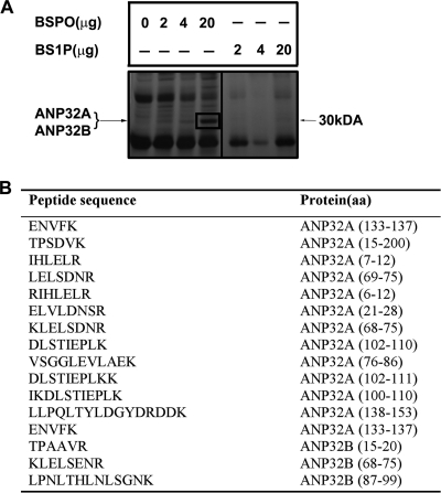
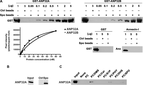
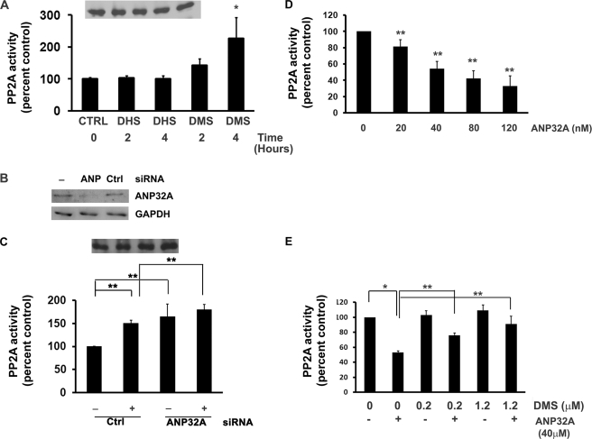
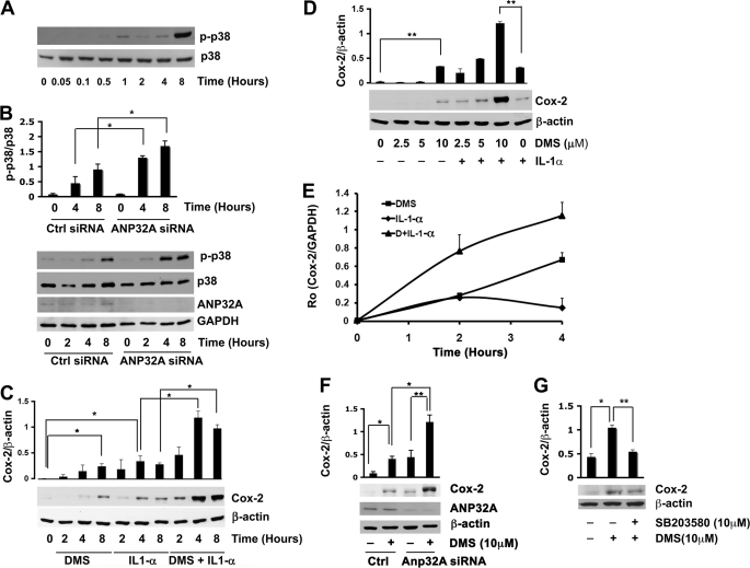
Sphingolipid metabolites regulate cell fate by acting on specific cellular targets. Although the influence of sphingolipids in cellular signaling has been well recognized, the exact molecular targets and how these targets influence cellular signaling mechanisms remain poorly understood. Toward this goal, we used affinity chromatography coupled with proteomics technology and identified acidic leucine-rich nuclear phosphoprotein-32A (ANP32A), an inhibitor of protein phosphatase 2A (PP2A) as a direct target of sphingosine, N,N'-dimethyl sphingosine (DMS) and phytosphingosine but not dihydrosphingosine or sphingosine 1-phosphate. Treatment of human umbilical vein endothelial cells (HUVEC) with DMS, which is not phosphorylated by sphingosine kinases, led to the activation of PP2A activity. Suppression of ANP32A with siRNA enhanced basal and DMS-activated PP2A activity suggesting that the sphingoid base binds to and relieves the inhibitory action of ANP32A on the PP2A complex. Indeed, DMS relieved the ANP32A-mediated inhibition of PP2A enzyme complex in vitro. Interestingly, DMS treatment induced the p38 stress-activated protein kinase (SAPK) and expression of cyclooxygenase (COX)-2 transcript and protein. Knockdown of ANP32A expression further induced p38 SAPK and COX-2. These data identify ANP32A as a novel molecular target of sphingoid bases that regulates cellular signaling events and inflammatory gene expression.


Figures

FIGURE 1.
FIGURE 1.
Identification of sphingosine-binding proteins. A, HEK293T whole cell extract (2 mg) was incubated for 1 h with BSPO or BS1P (2–20 μg) immobilized on a streptavidin membrane. After washing, BSPO, BS1P, or streptavidin membrane bound proteins were eluted with SDS sample buffer, separated by SDS-PAGE, and silver stained. The indicated band at 30 kDa representing proteins associated with BSPO was in-gel digested with trypsin and analyzed by LC-tandem mass spectrometry. B, 16 unique peptides that matched ANP32A and ANP32B are indicated.
FIGURE 2.
FIGURE 2.
Interaction of ANP32A and ANP32B with sphingolipids. A, different amounts of purified GST-ANP32B, GST-ANP32A, GST, or annexin I were incubated with sphingosine or control beads (1 mg) for 10 min, eluted with SDS sample buffer, and separated by SDS-PAGE. Western blot analysis was performed using an α-GST antibody or α-annexin I antibody. Band intensities were quantitated using a densitometer. B, HEK293T cell extract (1 mg) was incubated with sphingosine beads or control beads (1 mg), washed, eluted with SDS sample buffer, and separated by SDS-PAGE. Western blot analysis was performed using an α-ANP32A antibody. Input represents 50 μg of whole cell lysate. C, GST-ANP32A bound to indicated liposomes (250 μm) was centrifuged, and ANP32A present in the pellet was detected by immunoblot analysis.
FIGURE 3.
FIGURE 3.
DMS relieves the inhibitory action of ANP32A on PP2A. A, HUVEC were treated with 10 μm DMS or DHS for various times, and PP2A activity was determined as described under “Experimental Procedures.” PP2A was immunoprecipitated, and PP2A immunoblots are represented in the inset. Data represent mean ± S.D. of at least two independent experiments performed in duplicate. B, HUVEC were treated with ANP32A siRNA or control siRNA for 48 h. ANP32A and GAPDH protein levels were measured by immunoblot analysis. C, HUVEC were treated with ANP32A siRNA (80 nm) or control siRNA (80 nm) for 48 h, followed by treatment with DMS (10 μm) for 2 h or not and PP2A activity was determined. PP2A was immunoprecipitated, and PP2A immunoblots are represented in the inset. Data represent mean ± S.D. of at least two independent experiments performed in duplicate. D, PP2A dimer (containing A and C subunits) was incubated with increasing concentrations of ANP32A (0–120 nm), and its effects on PP2A activity was determined in vitro using the radioactive phosphatase assay, as described under “Experimental Procedures.” Data represent mean ± S.E. of at least three independent experiments performed in duplicate n = 6. E, purified ANP32A (40 nm) was incubated with various concentrations of DMS (0, 0.25, or 1.25 μm), and PP2A dimer activity were determined in vitro as described above. The data represent the mean ± S.E. of at least four independent experiments performed in duplicate n = 8.
FIGURE 4.
FIGURE 4.
DMS induces p38 stress-activated protein kinase and COX-2 expression in an ANP32A-dependent manner. A, HUVEC were treated with DMS for the indicated time times. Phospho-p38 SAPK and p38 SAPK protein levels were measured by immunoblot analysis. Results are represented as mean ± S.D. of at least two experiments. B, HUVEC were treated with ANP32A siRNA or control siRNA for 48 h followed by treatment with DMS (10 μm) for the indicated time intervals. Phospho-p38 SAPK, p38 SAPK, ANP32A, and GAPDH protein levels were measured by immunoblot analysis. Phospho-p38 SAPK protein levels were normalized to p38 SAPK protein levels. Results are represented as mean ± S.D. of at least two experiments. C, HUVEC were incubated with IL-1α (10 ng/ml), DMS (10 μm), or DMS in combination with IL-1α for the indicated time intervals. COX-2 protein levels normalized to β-actin protein levels were measured by immunoblot analysis. Results are represented as mean ± S.D. of at least two experiments. D, HUVEC cells were incubated with IL-1α (10 ng/ml), DMS, or DMS in combination with IL-1α for the concentrations of DMS indicated or remained untreated for 8 h. COX-2 protein levels normalized to β-actin protein levels were measured by immunoblot analysis. E, HUVEC were incubated with 10 μm DMS, 10 ng/ml IL-1α, or DMS and IL-1α for the indicated time intervals. COX-2 mRNA levels normalized to GAPDH mRNA levels were measured using qRT- PCR as described under “Experimental Procedures.” Results are represented as mean ± S.D. (n = 3). The experiment was repeated twice with similar results. F, HUVEC were treated with ANP32A siRNA or control siRNA for 48 h followed by treatment with DMS (10 μm) for 8 h. COX-2 protein levels normalized to β-actin protein levels were measured by immunoblot analysis. Results are represented as mean ± S.D. of at least two experiments. G, HUVEC cells were pretreated with SB203580 (10 μm) for 30 min, incubated with DMS (10 μm) for 8 h. COX-2 protein levels were measured by immunoblot analysis and normalized to β-actin levels. Results are expressed as means ± S.D. of at least two experiments. Results are represented as mean ± S.D. of at least two experiments. *, p ≤ 0.05; **, p ≤ 0.01 (two-tailed t test).

Similar articles

Cited by

References

    1. Bartke N., Hannun Y. A. (2009) J. Lipid Res. 50, (suppl.) S91–S96 - PMC - PubMed
    1. Hla T., Lee M. J., Ancellin N., Paik J. H., Kluk M. J. (2001) Science 294, 1875–1878 - PubMed
    1. Spiegel S., Milstien S. (2003) Nat. Rev. Mol. Cell Biol. 4, 397–407 - PubMed
    1. Morales A., Lee H., Goñi F. M., Kolesnick R., Fernandez-Checa J. C. (2007) Apoptosis 12, 923–939 - PubMed
    1. Cuvillier O. (2002) Biochim. Biophys. Acta 1585, 153–162 - PubMed

Publication types

MeSH terms