Thioredoxin-interacting protein (Txnip) gene expression: sensing oxidative phosphorylation status and glycolytic rate
- PMID: 20558747
- PMCID: PMC2919144
- DOI: 10.1074/jbc.M110.108290
Thioredoxin-interacting protein (Txnip) gene expression: sensing oxidative phosphorylation status and glycolytic rate
Abstract
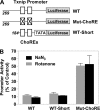
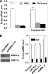
Thioredoxin-interacting protein (Txnip) has important functions in regulating cellular metabolism including glucose utilization; the expression of the Txnip gene is sensitive to the availability of glucose and other fuels. Here, we show that Txnip expression is down-regulated at the transcriptional level by diverse inhibitors of mitochondrial oxidative phosphorylation (OXPHOS). The effect of these OXPHOS inhibitors is mediated by earlier identified carbohydrate-response elements (ChoREs) on the Txnip promoter and the ChoRE-associated transcription factors Max-like protein X (MLX) and MondoA (or carbohydrate-response element-binding protein (ChREBP)) involved in glucose-induced Txnip expression, suggesting that inhibited oxidative phosphorylation compromises glucose-induced effects on Txnip expression. We also show that the OXPHOS inhibitors repress the Txnip transcription most likely by inducing the glycolytic rate, and increased glycolytic flux decreases the levels of glycolytic intermediates important for the function of MLX and MondoA (or ChREBP). Our findings suggest that the Txnip expression is tightly correlated with glycolytic flux, which is regulated by oxidative phosphorylation status. The identified link between the Txnip expression and glycolytic activity implies a mechanism by which the cellular glucose uptake/homeostasis is regulated in response to various metabolic cues, oxidative phosphorylation status, and other physiological signals, and this may facilitate our efforts toward understanding metabolism in normal or cancer cells.
Figures


Similar articles
-
Hypoxia-inducible factor independent down-regulation of thioredoxin-interacting protein in hypoxia.FEBS Lett. 2011 Feb 4;585(3):492-8. doi: 10.1016/j.febslet.2010.12.033. Epub 2010 Dec 27. FEBS Lett. 2011. PMID: 21192937
-
Tandem ChoRE and CCAAT motifs and associated factors regulate Txnip expression in response to glucose or adenosine-containing molecules.PLoS One. 2009 Dec 22;4(12):e8397. doi: 10.1371/journal.pone.0008397. PLoS One. 2009. PMID: 20027290 Free PMC article.
-
Glutamine-dependent anapleurosis dictates glucose uptake and cell growth by regulating MondoA transcriptional activity.Proc Natl Acad Sci U S A. 2009 Sep 1;106(35):14878-83. doi: 10.1073/pnas.0901221106. Epub 2009 Aug 17. Proc Natl Acad Sci U S A. 2009. PMID: 19706488 Free PMC article.
-
Interactions between Myc and MondoA transcription factors in metabolism and tumourigenesis.Br J Cancer. 2015 Dec 1;113(11):1529-33. doi: 10.1038/bjc.2015.360. Epub 2015 Oct 15. Br J Cancer. 2015. PMID: 26469830 Free PMC article. Review.
-
Glucose sensing by ChREBP/MondoA-Mlx transcription factors.Semin Cell Dev Biol. 2012 Aug;23(6):640-7. doi: 10.1016/j.semcdb.2012.02.007. Epub 2012 Mar 3. Semin Cell Dev Biol. 2012. PMID: 22406740 Review.
Cited by
-
Slow flow induces endothelial dysfunction by regulating thioredoxin-interacting protein-mediated oxidative metabolism and vascular inflammation.Front Cardiovasc Med. 2022 Nov 16;9:1064375. doi: 10.3389/fcvm.2022.1064375. eCollection 2022. Front Cardiovasc Med. 2022. PMID: 36465470 Free PMC article.
-
Purinergic regulation of high-glucose-induced caspase-1 activation in the rat retinal Müller cell line rMC-1.Am J Physiol Cell Physiol. 2011 Nov;301(5):C1213-23. doi: 10.1152/ajpcell.00265.2011. Epub 2011 Aug 10. Am J Physiol Cell Physiol. 2011. PMID: 21832250 Free PMC article.
-
Integrated analysis of transcriptional and metabolic responses to mitochondrial stress.Cell Rep Methods. 2025 Apr 21;5(4):101027. doi: 10.1016/j.crmeth.2025.101027. Epub 2025 Apr 14. Cell Rep Methods. 2025. PMID: 40233762 Free PMC article.
-
Thioredoxin-interacting protein gene expression via MondoA is rapidly and transiently suppressed during inflammatory responses.PLoS One. 2013;8(3):e59026. doi: 10.1371/journal.pone.0059026. Epub 2013 Mar 8. PLoS One. 2013. PMID: 23520550 Free PMC article.
-
Thioredoxin and thioredoxin target proteins: from molecular mechanisms to functional significance.Antioxid Redox Signal. 2013 Apr 1;18(10):1165-207. doi: 10.1089/ars.2011.4322. Epub 2012 Jun 26. Antioxid Redox Signal. 2013. PMID: 22607099 Free PMC article. Review.
References
-
- Kim S. Y., Suh H. W., Chung J. W., Yoon S. R., Choi I. (2007) Cell Mol. Immunol. 4, 345–351 - PubMed
-
- Chen C. L., Lin C. F., Chang W. T., Huang W. C., Teng C. F., Lin Y. S. (2008) Blood 111, 4365–4374 - PubMed
-
- Han S. H., Jeon J. H., Ju H. R., Jung U., Kim K. Y., Yoo H. S., Lee Y. H., Song K. S., Hwang H. M., Na Y. S., Yang Y., Lee K. N., Choi I. (2003) Oncogene 22, 4035–4046 - PubMed
Publication types
MeSH terms
Substances
LinkOut - more resources
Full Text Sources

