. 2010 Sep;109(3):820-9.
doi: 10.1152/japplphysiol.91430.2008. Epub 2010 Jun 17.

Hyperoxic stimulation of synchronous orthodromic activity and induction of neural plasticity does not require changes in excitatory synaptic transmission

Affiliations

Hyperoxic stimulation of synchronous orthodromic activity and induction of neural plasticity does not require changes in excitatory synaptic transmission

Alfredo J Garcia 3rd et al. J Appl Physiol (1985). 2010 Sep.

Abstract

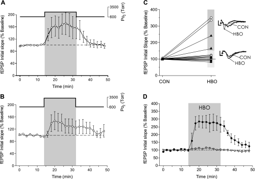
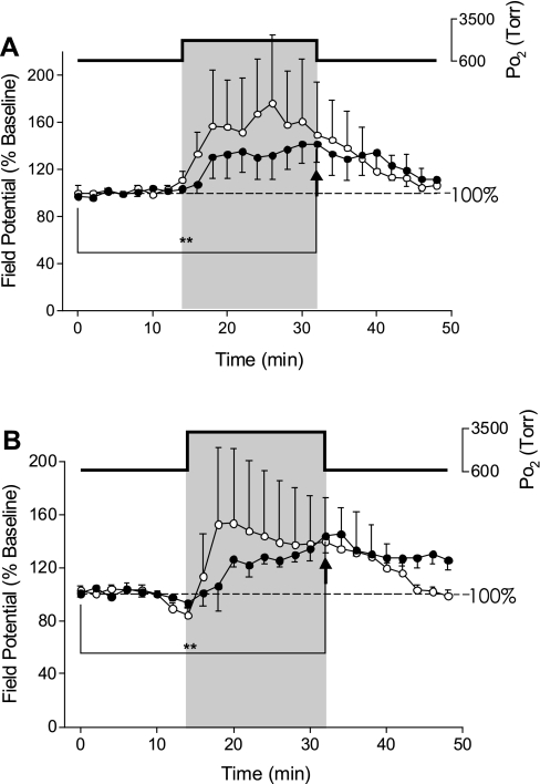
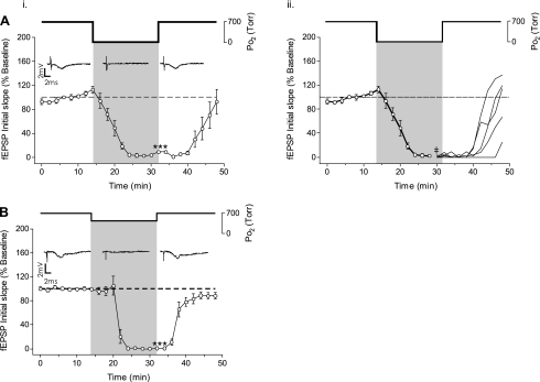
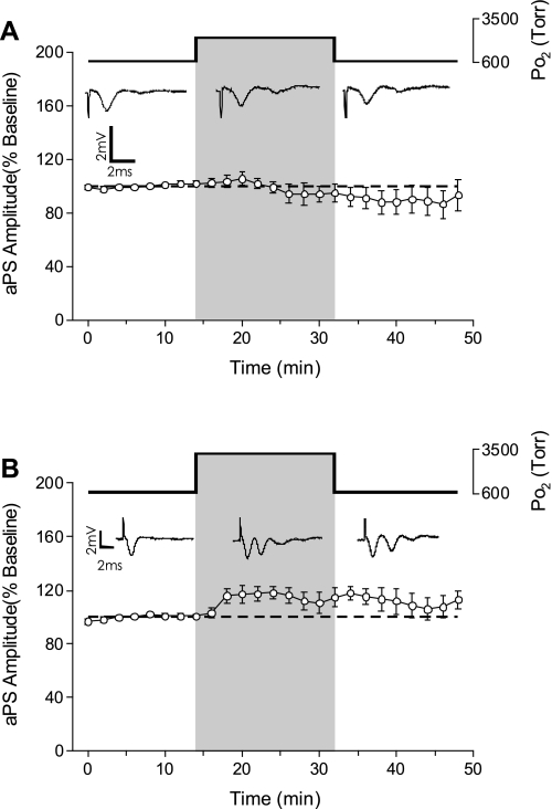
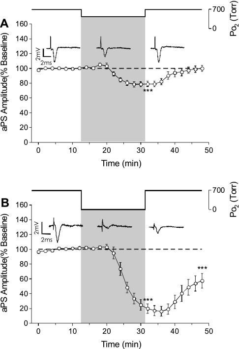
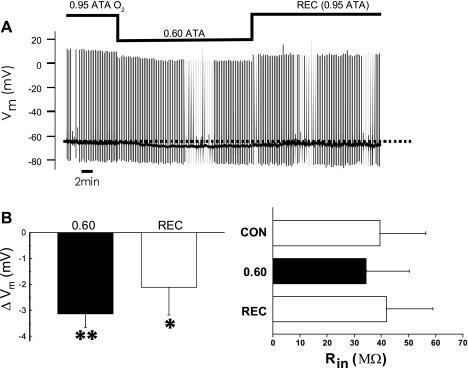
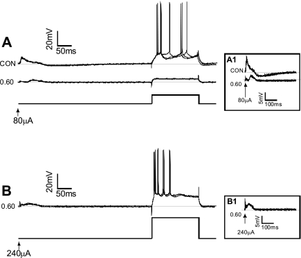
The first study, described in the companion article, reports that acute exposure of rat hippocampal slices to either hyperbaric oxygen (HBO: 2.84 and 4.54 atmospheres absolute, ATA) or normobaric reoxygenation (NBOreox; i.e., normobaric hyperoxia: 0.6 or 0.0→0.95 ATA) stimulates synchronous orthodromic activity in CA1 neurons, which includes activation of O2-induced potentiation (OxIP) and, in some cases, hyperexcitability (secondary population spikes, sPS). In this second study we tested the hypothesis that HBO and NBOreox increase orthodromic activity of CA1 neurons (oPS, orthodromic population spike) and OxIP via a combination of both increased excitatory synaptic transmission (field excitatory postsynaptic potential, fEPSP) and intrinsic excitability (antidromic population spike, aPS). HBO and NBOreox increased the oPS but rarely increased or potentiated the fEPSP. HBO exposure produced epileptiform antidromic activity, which was abolished during inhibition of fast GABAergic and glutamatergic synaptic transmission. Decreasing O2 from 0.95 ATA (control) to 0.6 ATA (intermediate O2) or 0.0 ATA (hypoxia) reversibly abolished the fEPSP, and reoxygenation rarely induced potentiation of the fEPSP or aPS. Intracellular recordings and antidromic field potential recordings, however, revealed that synaptic transmission and neuronal excitability were preserved, albeit at lower levels, in 0.60 ATA O2. Together, these data indicate that 1) the changes in excitatory postsynaptic activity are not required for stimulation of the oPS during and HBO/NBOreox or for activation of OxIP, suggesting the latter is a form of intrinsic plasticity; 2) HBO disinhibits spontaneous synaptic transmission to induce epileptiform activity; and 3) although synchronous synaptic activation of the CA1 neuronal population requires hyperoxia (i.e., 0.95 ATA O2), synaptic activation of individual CA1 neurons does not.


Figures

Fig. 1.
Fig. 1.
Effect of hyperbaric oxygen (HBO) on the field excitatory postsynaptic potential (fEPSP) in CA1 neurons during electrical stimulation of Schaffer collaterals. A: the fEPSP response in CA1 neurons during HBO was variable. Although a 16-min exposure to 2.84 ATA O2 appeared to stimulate the fEPSP, no significance was found in the averaged response to 2.84 ATA O2 exposure (n = 9). B: similar to the response to 2.84 ATA O2, the fEPSP was variable and not significantly affected during a 16-min exposure to 4.54 ATA O2 (n = 7). C: plotting the individual fEPSP experiments before (control, CON; t = 0 min) and during the exposure (HBO; t = 32 min) to either 2.84 ATA O2 (○) or 4.54 ATA O2 (▴) revealed that HBO stimulated the fEPSP in a subset of experiments (n = 4/16). Raw data traces are shown from experiments where HBO exposure stimulated the fEPSP ≥150% or where HBO did not significantly affect the fEPSP. The arrowheads denote the stimulus artifact removed from trace. D: the response to HBO could be divided into 2 groups: stimulation (●) or no effect (▿); however, the predominant fEPSP response to HBO was no effect (n = 12/16).
Fig. 2.
Fig. 2.
Effects of HBO on simultaneous recordings of the fEPSP and orthodromic population spike (oPS). A: exposure to 2.84 ATA O2 caused variable effects on the fEPSP (○). While the fEPSP was stimulated in only 1 slice, the oPS (●) was significantly (**P < 0.01) stimulated by exposure to 2.84 ATA O2 compared with baseline (n = 5) as denoted by the arrowhead. B: simultaneous recordings (n = 4) during exposure to 4.54 ATA O2 caused a significant stimulation (**P < 0.01) of the oPS (●), as denoted by the arrowhead, with a significant O2-induced potentiation (OxIP) on return to 0.95 ATA O2, but again, the fEPSP (○) was stimulated by HBO in only 1 of the 4 preparations. The oPS data at both levels of HBO were pooled with experiments recording just the oPS as reported in Ref. .
Fig. 3.
Fig. 3.
Effect of 0.60 and 0.00 ATA O2 on the fEPSP. Ai: exposure to 16 min of 0.00 ATA O2 significantly inhibited the fEPSP (n = 5, ***P > 0.001). The fEPSP recovered to baseline within 16 min on return to the control 0.95 ATA O2. ii: the mean recovery response of the fEPSP from 0.00ATA O2 (i) was separated into the individual recoveries (beginning at ‡) showing that recovery of the fEPSP from hypoxia was variable, with some (n = 2/5) preparations not recovering to baseline and others overshooting baseline (OxIP) by the end of the 16-min recovery period (t = 48 min). B: exposure to 16 min of 0.60 ATA O2 also significantly inhibited the fEPSP (n = 5, ***P > 0.001), which recovered to baseline within 16 min on return to the control 0.95 ATA O2 with no preparation exhibiting an overshoot of baseline activity.
Fig. 4.
Fig. 4.
Effects of HBO on the antidromic population spike (aPS). A: a 16-min exposure to 2.84 ATA O2 did not affect the amplitude of the aPS (n = 6). B: a 16-min exposure to 4.54 ATA O2 likewise did not significantly stimulate the amplitude of the aPS (n = 6). However, note the occurrence of secondary population spikes (sPS) during and following exposure to 4.54 ATA O2 (n = 3/6).
Fig. 5.
Fig. 5.
Synaptic blockade medium prevents the occurrence of sPS induced during and following exposure to 4.54 ATA O2. A: in the presence of synaptic blockade (50 μM picrotoxin, 100 μM MK-801, 50 and 20 μM CNQX), exposure to 4.54 ATA O2 had no effect on the aPS amplitude, but the occurrence of sPS were eliminated (n = 7). Arrowhead denotes stimulus artifact removed from the raw data trace. B: exposure to the GABAA receptor antagonist picrotoxin (50–65 μM) alone while maintaining medium O2 tension (PmO2) constant at 0.95 ATA O2 caused no significant change in the aPS but consistently produced sPS (asterisk) in all slices tested during exposure and recovery (n = 5). Arrowhead denotes stimulus artifact removed from the raw data trace.
Fig. 6.
Fig. 6.
Effects of normobaric oxygen manipulation on the aPS. A: exposure to 0.60 ATA O2 caused a significant suppression in the aPS that was reversible upon return to control O2 (n = 7, ***P < 0.001). B: exposure to 0.00 ATA O2 caused a significant suppression in the aPS that was greater than the inhibition during exposure to 0.60 ATA O2 (n = 7, ***P < 0.001). On return to 0.95 ATA O2, the aPS did not return to its baseline level but continued to be suppressed (***P < 0.001).
Fig. 7.
Fig. 7.
Effects of intermediate levels of O2 on the membrane potential (Vm) and input resistance (Rin) of individual CA1 neurons. The rationale for recording Vm during acute exposure to 0.60 ATA O2 was to determine whether CA1 neurons remained electrophysiologically viable during intermediate oxygenation, since orthodromic field potential recordings (oPS, fEPSP) suggested that they did not, but antidromic field potential recordings (aPS) suggested that they did. A: raw data of a nonspontaneously firing CA1 neuron. Action potentials could be evoked with current injection, whereas excitatory postsynaptic potentials (EPSPs) could be evoked with extracellular stimulation. To evoke action potentials and EPSPs during intermediate oxygenation, the amount of current injected intracellularly or extracellularly had to be increased (see Fig. 8). The trace also illustrates the hyperpolarization below the mean Vm value under initial control conditions (dashed line) without a measurable change in Rin. B, left: summary bar graph showing that exposure to 0.60 ATA O2 caused a reversible (P > 0.05) hyperpolarization that was significantly different from control as tested by a one-sample t-test (**P < 0.05) during exposure to 0.60 ATA O2 (n = 5). Right, summary bar graph showing Rin was not significantly altered on average during or following (P > 0.05) exposure to 0.60 ATA O2 (n = 5). Rec, recovery.
Fig. 8.
Fig. 8.
Effects of intermediate level of O2 on the action potential generation and the evoked EPSP of individual CA1 neurons measured during intracellular recording. The rationale for these experiments was to determine whether CA1 neurons retained the capacity for synaptic activation during acute exposure to 0.6 ATA O2, since orthodromic field potential recordings suggested that they did not. A: action potentials and EPSPs could be evoked by depolarizing current and extracellular stimulation of Schaffer collaterals under control conditions (0.95 ATA O2). on exposure to 0.60 ATA O2, the same depolarizing current protocol could not evoke action potentials, whereas the extracellular stimulating current could only intermittently evoke attenuated EPSPs. In this example, Rin decreased during exposure to 0.6 ATA O2; however, the change in Rin during exposure to intermediate oxygenation, on average, was not significant (Fig. 7B). Inset A1: high-gain images of the evoked EPSPs under control and 0.60 ATA O2 conditions. B: by increasing the amount of depolarizing current injected, it was possible to evoke repetitive firing of action potentials in 0.60 ATA O2. Moreover, increasing the extracellular stimulation current also increased the amplitude of the EPSP, although it was still attenuated compared with the EPSPs generated under control O2 conditions. Inset B1: high-gain images of an EPSP generated in 0.60 ATA O2 using 3× the stimulation current in the same cell as shown in A.
Fig. 9.
Fig. 9.
Mean responses of field potentials as a function of measured PmO2 at normobaric pressure (NBO) and HBO. Mean responses of oPS, fEPSP, and aPS are plotted as a function of PmO2. The oPS data are reproduced from Fig. 9A in the first of the companion articles (9). Multiple measures of CA1 neuronal excitability indicate that hippocampal slices maintained in normobaric hyperoxia (0.95 ATA O2) exhibit their greatest O2 sensitivity in the range of normobaric oxygenation (NBO: oPS, fEPSP, and aPS). In contrast, O2 sensitivity of the mechanisms underlying the aPS and fEPSP plateaus are saturated beginning at 0.95 ATA O2. Likewise, the O2 sensitivity of the oPS is significantly reduced beginning at 0.95 ATA O2 and above (see Ref. for statistics).
Fig. 10.
Fig. 10.
Working model derived from our two studies (Ref. and present study) that postulates how acute exposure to HBO may stimulate CA1 neurons causing increased excitability and OxIP. This model may also be applied to the phenomenon of OxIP of the oPS caused by NBOreox from 0.60 to 0.95 ATA O2. A: in 0.95 ATA O2 (control conditions), excitability of CA1 pyramidal neurons (open and shaded bars) is regulated by active synaptic input from GABAergic interneurons [open box, inhibitory (−) neurons] and from Schaffer-commissural collaterals and intrinsic membrane properties of the pyramidal neurons themselves. Moreover, with submaximal stimulation of the Schaffer-commissural collaterals, as used in our studies, only a fraction of neurons are activated (open), whereas others are silent (shaded). B: during HBO, our study shows that the oPS is stimulated (1), and thus more CA1 pyramidal neurons are activated with Schaffer-commissural collateral stimulation. The elevated excitability of CA1 neurons during HBO is not dependent on a significant increase in the excitatory synapses of the Schaffer-commissural collaterals as measured by the lack of significant change on average of the fEPSP (2) or lack of change in axonal conduction as measured by the primary aPS (3). Finding that onset of antidromic sPS (3) during and following HBO exposure could be blocked by chemical synaptic blockade and mimicked by the application of picrotoxin (Ptx), which inhibits GABAergic ionotropic receptors, suggests that epileptiform activity occurs through GABAergic interneurons (shaded box) (4). The lack of effect of HBO on the primary aPS and fEPSP suggests that elevated excitability of CA1 pyramidal neurons results from a change in conduction from synapse to soma (5). Although it has yet to be determined, potential targets of O2 (and presumably ROS/RNS) that would increase orthodromic activation and induction of OxIP of the oPS include voltage-gated channels that regulate dendritic conduction and the threshold for action potential generation and gap junctions (shaded area).

Similar articles

Cited by

References

    1. Bitterman N, Halpern P. The effect of flumazenil on CNS oxygen toxicity in the rat. Methods Find Exp Clin Pharmacol 17: 169–174, 1995 - PubMed
    1. Brahman B, Forman RE, Stewart EE, Nicholson C, Rice ME. Ascorbate inhibits edema in brain slices. J Neurochem 74: 1263–1270, 2000 - PubMed
    1. D'Agostino DP, Putnam RW, Dean JB. Superoxide (•O2−) production in CA1 neurons of rat hippocampal slices exposed to graded levels of oxygen. J Neurophysiol 98: 1030–1041, 2007 - PubMed
    1. Daoudal G, Dubane D. Long-term plasticity of intrinsic excitability: learning rules and memory. Learn Mem 10: 456–465, 2003 - PubMed
    1. Dean J. Hypercapnia causes cellular oxidation and nitrosation in addition to acidosis: implications for CO2 chemoreceptor function and dysfunction. J Appl Physiol 108: 1786–1795, 2010 - PMC - PubMed

Publication types

MeSH terms