. 2010 Jul 23;107(2):294-304.
doi: 10.1161/CIRCRESAHA.110.223172. Epub 2010 Jun 17.

Myocardin-related transcription factor-a controls myofibroblast activation and fibrosis in response to myocardial infarction

Affiliations

Myocardin-related transcription factor-a controls myofibroblast activation and fibrosis in response to myocardial infarction

Eric M Small et al. Circ Res. .

Abstract

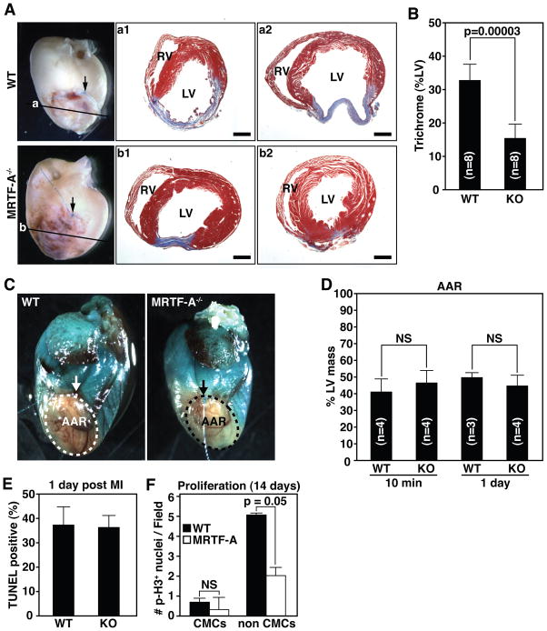
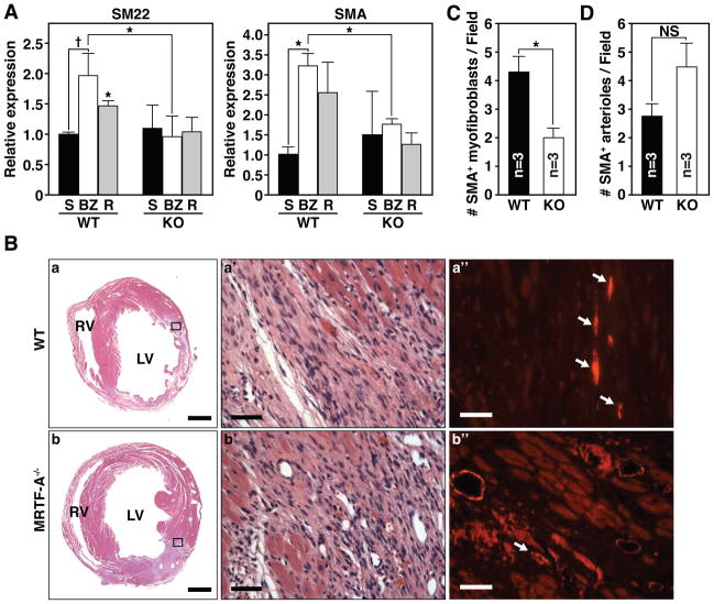
Rationale: Myocardial infarction (MI) results in loss of cardiac myocytes in the ischemic zone of the heart, followed by fibrosis and scar formation, which diminish cardiac contractility and impede angiogenesis and repair. Myofibroblasts, a specialized cell type that switches from a fibroblast-like state to a contractile, smooth muscle-like state, are believed to be primarily responsible for fibrosis of the injured heart and other tissues, although the transcriptional mediators of fibrosis and myofibroblast activation remain poorly defined. Myocardin-related transcription factors (MRTFs) are serum response factor (SRF) cofactors that promote a smooth muscle phenotype and are emerging as components of stress-responsive signaling.

Objective: We aimed to examine the effect of MRTF-A on cardiac remodeling and fibrosis.

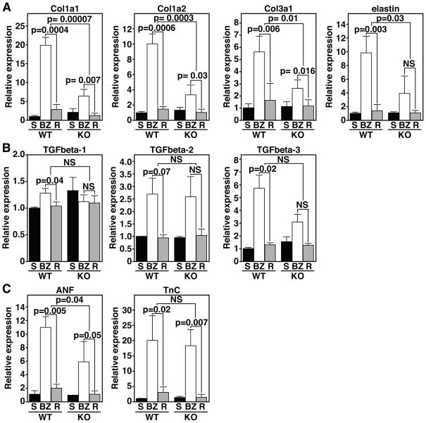
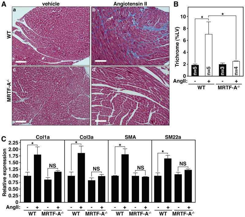
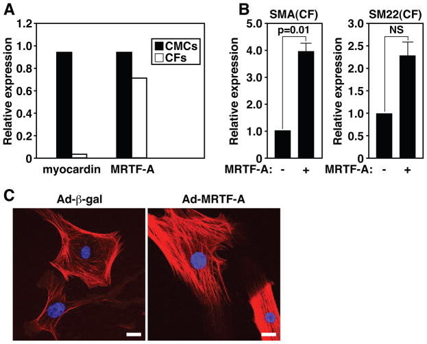
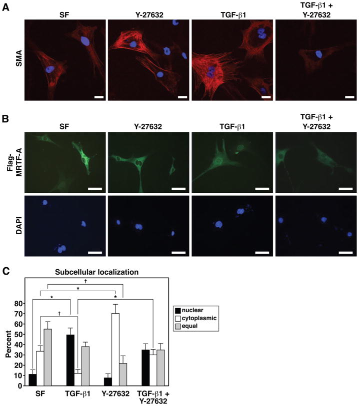
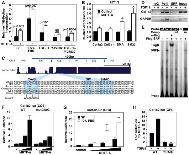
Methods and results: Here, we show that MRTF-A controls the expression of a fibrotic gene program that includes genes involved in extracellular matrix production and smooth muscle cell differentiation in the heart. In MRTF-A-null mice, fibrosis and scar formation following MI or angiotensin II treatment are dramatically diminished compared with wild-type littermates. This protective effect of MRTF-A deletion is associated with a reduction in expression of fibrosis-associated genes, including collagen 1a2, a direct transcriptional target of SRF/MRTF-A.

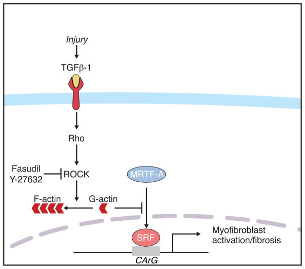
Conclusions: We conclude that MRTF-A regulates myofibroblast activation and fibrosis in response to the renin-angiotensin system and post-MI remodeling.


Figures

Figure 1
Figure 1. MRTF-A deletion results in reduced scar formation following MI
(A) Whole-mount images of representative hearts 14 days post-MI and Masson’s trichrome staining of sections from two representative hearts. Arrow denotes point of ligature and plane of section is illustrated by horizontal line. Masson’s trichrome staining illustrates reduced and more compact region of scarring in the infarct zone (compare a1 and a2 to b1 and b2) RV, right ventricle; LV, left ventricle. Scale bar = 1 mm. (B) Quantification of infarct size presented as the percent LV area positively stained with Masson’s trichrome. n=8 WT and 8 KO animals. (C) Area at risk (AAR) determined by perfusion with methylene blue. AAR is devoid of staining. Arrow denotes point of ligature on the LAD. (D) Quantification of AAR presented as percent of LV mass that is not stained 10 minutes and 1-day post-MI. n=4 WT and 4 MRTF-A−/− animals at 10 min and 3 WT and 3 MRTF-A−/− animals 1-day post-MI. (E) Quantification of TUNEL positive cells 1 day post-MI was performed on at least 4 independent fields within the infarct zone of each heart and averaged from 2 WT and 3 MRTF-A−/− animals. Data is represented as percentage of Dapi stained nuclei positive for TUNEL. (F) Quantification of phospho-histone H3 postive cells 14 days post-MI was performed on at least 7 independent fields in the BZ from each heart and averaged from 3 WT and 4 MRTF-A−/− animals. Error bars indicate the SEM.
Figure 2
Figure 2. MRTF-A influences ECM gene expression 14 days post-MI
(A) Real time PCR for various ECM components in sham hearts (S) and the border zone (BZ) or remote (R) region of the infarct reveals attenuated expression of collagen genes in the BZ of MRTF-A−/− hearts. (B) The cytokines TGFβ-1, -2, and -3 do not display significant alterations in expression in the BZ between WT and MRTF-A−/− hearts. (C) The stress responsive ANF gene displays reduced expression in MRTF-A−/− hearts. TnC levels are elevated to similar levels in the BZ of both WT and MRTF-A−/− animals. n=4 WT and 4 MRTF-A−/− hearts for infarct groups and 2 shams. Error bars represent the SEM.
Figure 3
Figure 3. MRTF-A deletion results in altered smooth muscle gene expression 14 days post-MI
(A) Real time PCR reveals a significant reduction of SM22 and SMA induction in both the BZ and remote region of MRTF-A−/− hearts (* denotes p-value < 0.01, and † denotes p-value < 0.05). n=4 WT and 4 MRTF-A−/− hearts per infarct group and 2 shams. (B) Histological sections of hearts from WT and MRTF-A−/− mice, 14 days post-MI are shown. H&E stained (a, b) sections of representative hearts adjacent to those used for immunohistochemistry illustrate ischemic damage and highlight the BZ of the infarct. (a’ and b’) Magnification of region that is boxed in a and b. (a” and b”) SMA immunostaining corresponding to the boxed region of representative WT and MRTF-A−/− infarcted hearts. Arrows mark SMA-positive spindle-shaped myofibroblasts. Scale bar for a, b = 1mm. Scale bar for a’, a” and b’, b” = 40 μm. (C) Number of SMA-positive myofibroblasts per field of view. (D) Number of SMA positive arterioles in the BZ per field of view. n=3 WT and 3 MRTF-A−/− hearts. Error bars represent the SEM. Quantification was performed at 40x magnification on at least 3 fields of view within the BZ of each heart and averaged from 3 WT and 3 MRTF-A−/− hearts (* denotes p-value < 0.05). Error bars represent the SEM.
Figure 4
Figure 4. MRTF-A deletion attenuates fibrosis following chronic AngII administration
(A) Masson’s trichrome staining of representative histological sections following 14 days of vehicle (a, c) or AngII (b, d) administration in WT (a, b) or MRTF-A−/− (c, d) mice. (B) Quantification of trichrome staining of the left ventricle (LV). Error bars represent SEM (* denotes p-value < 0.05). (C) Quantitative RT-PCR for collagen genes and smooth muscle differentiation markers reveals attenuated induction of Col1a, Col3a, SMA and SM22 following AngII administration of MRTF-A−/− animals. Error bars represent SEM (* denotes p-value < 0.05).
Figure 5
Figure 5. MRTF-A regulates the expression of smooth muscle markers in CFs
(A) Quantitative Real Time PCR reveals myocardin is exclusively expressed in CMCs, while MRTF-A is present in both CMCs and CFs at similar levels. (B) Quantitative Real Time RT-PCR reveals SMA and SM22 are significantly enriched in CFs over-expressing MRTF-A relative to β-gal infected control CFs. Error bars represent the SEM. (C) Indirect immunofluorescence of CFs demonstrates MRTF-A over-expression results in the enrichment of SMA into organized stress fibers as compared with Ad-β-gal infected CFs, which display primarily cortical actin SMA staining. Scale bar = 25 μm.
Figure 6
Figure 6. Regulation of MRTF-A nuclear localization and activity in CFs by TGFβ-1 and the ROCK inhibitor, Y-27632
(A) Immunocytochemistry for SMA in CFs grown in serum free (SF) media or media supplemented with TGFβ-1 (10ng/ml), Y-27632 (10μM) or both TGFβ-1 and Y-27632. All images were captured using identical exposure settings. Scale bar = 25 μm. (B) Immunocytochemical detection of Flag-tagged MRTF-A in CFs after 24 hours of growth in SF media or media supplemented with TGFβ-1, Y-27632 or both TGFβ-1 and Y-27632. Scale bar = 100 μm. (C) Quantification of the subcellular localization of Flag-MRTF-A under various culture conditions. Subcellular localization of Flag-MRTF-A was scored for approximately 20 random fields of view for each condition. Error bars represent the SEM (* denotes p-value < 0.01; † denotes p-value < 0.05).
Figure 7
Figure 7. MRTF-A regulates Col1a2 expression
(A) [3H]-proline incorporation in CFs demonstrates MRTF-A significantly enriches collagen synthesis. CFs were serum starved for 48 hrs and then infected with 10 MOI Ad-MRTF-A or Ad-β-gal and cultured in SF media or media supplemented with 2.5% FBS, TGFβ-1 (10 ng/ml), Y-27632 (10 μM), or both TGFβ-1 and Y-27632 for an additional 48 hrs. Error bars represent SEM. (B) Quantitative Real Time RT-PCR demonstrating the expression of Col1a2, Col3a1, SMA and SM22 in 10T1/2 cells transfected with empty vector control, or MRTF-A expression vector. (C) Depiction of the region used in transient transfection assays and EMSAs are shown. Blue peaks denote evolutionary conservation and alignment of mammalian regulatory sequences are at bottom highlighting the conserved CArG, SP1 and Smad sites. CArG mutation used in EMSA and transient transfection assays is aligned with WT CArG. (D) Chromatin immunoprecipitation of Col1a2 or GAPDH promoter sequences with an antibody directed against endogenous SRF. Antibodies directed against PolII or IgG serve as positive and negative controls. Input is 1% of total chromatin. (E) Electrophoretic mobility assay demonstrates that SRF binds to the conserved CArG box. Flag antibody supershifts the SRF/DNA complex, while WT unlabeled competitor oligonucleotide abolishes the shifted complex. Mutant oligonucleotide (m) fails to bind SRF and unlabeled mutant competitor (m) fails to abolish SRF/CArG interaction. (F) Transient transfection of COS cells with a WT or CArG mutant Col1a2-luciferase construct reveals dose-dependent responsiveness to MRTF-A. Error bars represent the SD. (G) MRTF-A induces the expression of Col1a2-luc in transiently transfected CFs and displays increased activity in the presence of 10% FBS. Error bars represent the SD. (H) MRTF-A dependent Col1a2 promoter activity is stimulated by TGFβ-1 treatment. Mutation of the CArG box abrogates responsiveness of the promoter to MRTF-A or TGFβ-1 treatment. Error bars represent the SD.
Figure 8
Figure 8. Proposed mechanism of MRTF-A activity during the stress response to MI
Increased TGFβ-1 levels result in the nuclear accumulation of MRTF-A in a Rho-ROCK dependent manner. Nuclear MRTF-A targets CArG box containing genes and induces the expression of smooth muscle and ECM molecules indicative of a myofibroblast cell type.

Comment in

References

    1. Swynghedauw B. Molecular mechanisms of myocardial remodeling. Physiol Rev. 1999;79:215–262. - PubMed
    1. Tomasek JJ, Gabbiani G, Hinz B, Chaponnier C, Brown RA. Myofibroblasts and mechano-regulation of connective tissue remodelling. Nat Rev Mol Cell Biol. 2002;3:349–363. - PubMed
    1. Serini G, Gabbiani G. Mechanisms of myofibroblast activity and phenotypic modulation. Exp Cell Res. 1999;250:273–283. - PubMed
    1. Wang J, Chen H, Seth A, McCulloch CA. Mechanical force regulation of myofibroblast differentiation in cardiac fibroblasts. Am J Physiol Heart Circ Physiol. 2003;285:H1871–1881. - PubMed
    1. Hinz B. Formation and function of the myofibroblast during tissue repair. J Invest Dermatol. 2007;127:526–537. - PubMed

Publication types

MeSH terms