

Diversity in the molecular and cellular strategies of epithelium-to-mesenchyme transitions: Insights from the neural crest
- PMID: 20559020
- PMCID: PMC2958624
- DOI: 10.4161/cam.4.3.12501
Diversity in the molecular and cellular strategies of epithelium-to-mesenchyme transitions: Insights from the neural crest
Abstract
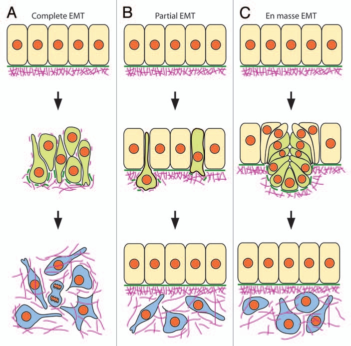
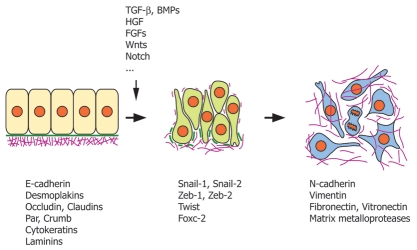
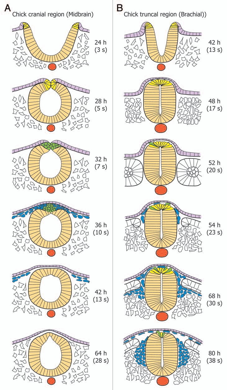
Although epithelial to mesenchymal transitions (EMT) are often viewed as a unique event, they are characterized by a great diversity of cellular processes resulting in strikingly different outcomes. They may be complete or partial, massive or progressive, and lead to the complete disruption of the epithelium or leave it intact. Although the molecular and cellular mechanisms of EMT are being elucidated owing chiefly from studies on transformed epithelial cell lines cultured in vitro or from cancer cells, the basis of the diversity of EMT processes remains poorly understood. Clues can be collected from EMT occuring during embryonic development and which affect equally tissues of ectodermal, endodermal or mesodermal origins. Here, based on our current knowledge of the diversity of processes underlying EMT of neural crest cells in the vertebrate embryo, we propose that the time course and extent of EMT do not depend merely on the identity of the EMT transcriptional regulators and their cellular effectors but rather on the combination of molecular players recruited and on the possible coordination of EMT with other cellular processes.
Figures


Similar articles
-
Sip1 mediates an E-cadherin-to-N-cadherin switch during cranial neural crest EMT.J Cell Biol. 2013 Dec 9;203(5):835-47. doi: 10.1083/jcb.201305050. Epub 2013 Dec 2. J Cell Biol. 2013. PMID: 24297751 Free PMC article.
-
MMP14 Regulates Cranial Neural Crest Epithelial-to-Mesenchymal Transition and Migration.Dev Dyn. 2018 Sep;247(9):1083-1092. doi: 10.1002/dvdy.24661. Epub 2018 Sep 9. Dev Dyn. 2018. PMID: 30079980 Free PMC article.
-
Cadherin-6B proteolysis promotes the neural crest cell epithelial-to-mesenchymal transition through transcriptional regulation.J Cell Biol. 2016 Dec 5;215(5):735-747. doi: 10.1083/jcb.201604006. Epub 2016 Nov 17. J Cell Biol. 2016. PMID: 27856599 Free PMC article.
-
Epithelial to mesenchymal transition during mammalian neural crest cell delamination.Semin Cell Dev Biol. 2023 Mar 30;138:54-67. doi: 10.1016/j.semcdb.2022.02.018. Epub 2022 Mar 8. Semin Cell Dev Biol. 2023. PMID: 35277330 Review.
-
"Beyond transcription: How post-transcriptional mechanisms drive neural crest EMT".Genesis. 2024 Feb;62(1):e23553. doi: 10.1002/dvg.23553. Epub 2023 Sep 21. Genesis. 2024. PMID: 37735882 Free PMC article. Review.
Cited by
-
A potential inhibitory function of draxin in regulating mouse trunk neural crest migration.In Vitro Cell Dev Biol Anim. 2017 Jan;53(1):43-53. doi: 10.1007/s11626-016-0079-0. Epub 2016 Sep 20. In Vitro Cell Dev Biol Anim. 2017. PMID: 27649978 Free PMC article.
-
Thrombospondin 1 promotes an aggressive phenotype through epithelial-to-mesenchymal transition in human melanoma.Oncotarget. 2014 Jul 30;5(14):5782-97. doi: 10.18632/oncotarget.2164. Oncotarget. 2014. PMID: 25051363 Free PMC article.
-
Investigating epithelial-to-mesenchymal transition with integrated computational and experimental approaches.Phys Biol. 2019 Mar 7;16(3):031001. doi: 10.1088/1478-3975/ab0032. Phys Biol. 2019. PMID: 30665206 Free PMC article. Review.
-
Dissection, Culture and Analysis of Primary Cranial Neural Crest Cells from Mouse for the Study of Neural Crest Cell Delamination and Migration.J Vis Exp. 2019 Oct 3;(152):10.3791/60051. doi: 10.3791/60051. J Vis Exp. 2019. PMID: 31633677 Free PMC article.
-
Contribution of neural crest-derived stem cells and nasal chondrocytes to articular cartilage regeneration.Cell Mol Life Sci. 2020 Dec;77(23):4847-4859. doi: 10.1007/s00018-020-03567-y. Epub 2020 Jun 5. Cell Mol Life Sci. 2020. PMID: 32504256 Free PMC article. Review.
References
-
- Stoker M, Gherardi E, Perryman M, Gray J. Scatter factor is a fibroblast-derived modulator of epithelial cell mobility. Nature. 1987;327:3–8. - PubMed
-
- Thiery JP, Acloque H, Huang RY, Nieto MA. Epithelialmesenchymal transitions in development and disease. Cell. 2009;139:871–890. - PubMed
-
- Thiery JP. Epithelial-mesenchymal transitions in tumour progression. Nat Rev Cancer. 2002;2:442–454. - PubMed
MeSH terms
LinkOut - more resources
Full Text Sources