ZAK is required for doxorubicin, a novel ribotoxic stressor, to induce SAPK activation and apoptosis in HaCaT cells
- PMID: 20559024
- PMCID: PMC3040836
- DOI: 10.4161/cbt.10.3.12367
ZAK is required for doxorubicin, a novel ribotoxic stressor, to induce SAPK activation and apoptosis in HaCaT cells
Abstract
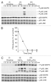
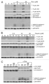
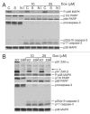
Doxorubicin is an anthracycline drug that is one of the most effective and widely used anticancer agents for the treatment of both hematologic and solid tumors. The stress-activated protein kinases (SAPKs) are frequently activated by a number of cancer chemotherapeutics. When phosphorylated, the SAPKs initiate a cascade that leads to the production of proinflammatory cytokines. Some inhibitors of protein synthesis, known as ribotoxic stressors, coordinately activate SAPKs and lead to apoptotic cell death. We demonstrate that doxorubicin effectively inhibits protein synthesis, activates SAPKs, and causes apoptosis. Ribotoxic stressors share a common mechanism in that they require ZAK, an upstream MAP3K, to activate the pro-apoptotic and proinflammatory signaling pathways that lie downstream of SAPKs. By employing siRNA mediated knockdown of ZAK or administration of sorafenib and nilotinib, kinase inhibitors that have a high affinity for ZAK, we provide evidence that ZAK is required for doxorubicin-induced proinflammatory and apoptotic responses in HaCaT cells, a pseudo-normal keratinocyte cell line, but not in HeLa cells, a cancerous cell line. ZAK has two different isoforms, ZAK-α (91 kDa) and ZAK-β (51 kDa). HaCaT or HeLa cells treated with doxorubicin and immunoblotted for ZAK displayed a progressive decrease in the ZAK-α band and the appearance of ZAK-β bands of larger size. Abrogation of these changes after exposure of cells to sorafenib and nilotinib suggests that these alterations occur following stimulation of ZAK. We suggest that ZAK inhibitors such as sorafenib or nilotinib may be effective when combined with doxorubicin to treat cancer patients.
Figures


Similar articles
-
Inhibition of doxorubicin-induced autophagy in hepatocellular carcinoma Hep3B cells by sorafenib--the role of extracellular signal-regulated kinase counteraction.FEBS J. 2011 Sep;278(18):3494-507. doi: 10.1111/j.1742-4658.2011.08271.x. FEBS J. 2011. PMID: 21790999
-
Small molecule kinase inhibitors block the ZAK-dependent inflammatory effects of doxorubicin.Cancer Biol Ther. 2013 Jan;14(1):56-63. doi: 10.4161/cbt.22628. Epub 2012 Oct 31. Cancer Biol Ther. 2013. PMID: 23114643 Free PMC article.
-
The flip side of doxorubicin: Inflammatory and tumor promoting cytokines.Cancer Biol Ther. 2013 Sep;14(9):774-5. doi: 10.4161/cbt.26125. Epub 2013 Aug 12. Cancer Biol Ther. 2013. PMID: 23974349 Free PMC article.
-
ZAK: a MAP3Kinase that transduces Shiga toxin- and ricin-induced proinflammatory cytokine expression.Cell Microbiol. 2008 Jul;10(7):1468-77. doi: 10.1111/j.1462-5822.2008.01139.x. Epub 2008 Mar 10. Cell Microbiol. 2008. PMID: 18331592
-
Sorafenib suppresses JNK-dependent apoptosis through inhibition of ZAK.Mol Cancer Ther. 2014 Jan;13(1):221-9. doi: 10.1158/1535-7163.MCT-13-0561. Epub 2013 Oct 29. Mol Cancer Ther. 2014. PMID: 24170769 Free PMC article.
Cited by
-
Ribosomal stress-surveillance: three pathways is a magic number.Nucleic Acids Res. 2020 Nov 4;48(19):10648-10661. doi: 10.1093/nar/gkaa757. Nucleic Acids Res. 2020. PMID: 32941609 Free PMC article. Review.
-
Mixed lineage kinase ZAK promotes epithelial-mesenchymal transition in cancer progression.Cell Death Dis. 2018 Feb 2;9(2):143. doi: 10.1038/s41419-017-0161-x. Cell Death Dis. 2018. PMID: 29396440 Free PMC article.
-
Emerging Role of GCN1 in Disease and Homeostasis.Int J Mol Sci. 2024 Mar 5;25(5):2998. doi: 10.3390/ijms25052998. Int J Mol Sci. 2024. PMID: 38474243 Free PMC article. Review.
-
Cell cycle regulation by the ribotoxic stress response.Trends Cell Biol. 2025 Jul;35(7):592-603. doi: 10.1016/j.tcb.2025.04.005. Epub 2025 May 16. Trends Cell Biol. 2025. PMID: 40379527 Review.
-
Nilotinib protects the murine liver from ischemia/reperfusion injury.J Hepatol. 2012 Oct;57(4):766-73. doi: 10.1016/j.jhep.2012.05.012. Epub 2012 May 26. J Hepatol. 2012. PMID: 22641092 Free PMC article.
References
-
- Takemura G, Fujiwara H. Doxorubicin-induced cardiomyopathy from the cardiotoxic mechanisms to management. Prog Cardiovasc Dis. 2007;49:330–352. - PubMed
-
- Gewirtz DA. A critical evaluation of the mechanisms of action proposed for the antitumor effects of the anthracycline antibiotics adriamycin and daunorubicin. Biochem Pharmacol. 1999;57:727–741. - PubMed
-
- Singal PK, Li T, Kumar D, Danelisen I, Iliskovic N. Adriamycin-induced heart failure: mechanism and modulation. Mol Cell Biochem. 2000;207:77–86. - PubMed
-
- Wood LJ, Nail LM, Gilster A, Winters KA, Elsea CR. Cancer chemotherapy-related symptoms: evidence to suggest a role for proinflammatory cytokines. Oncol Nurs Forum. 2006;33:535–542. - PubMed
-
- Kalyanaraman B, Joseph J, Kalivendi S, Wang S, Konorev E, Kotamraju S. Doxorubicin-induced apoptosis: implications in cardiotoxicity. Mol Cell Biochem. 2002;234:119–124. - PubMed
Publication types
MeSH terms
Substances
Grants and funding
LinkOut - more resources
Full Text Sources
Molecular Biology Databases
