Review
. 2010 Jul;6(7):489-97.
doi: 10.1038/nchembio.392.

An update on sphingosine-1-phosphate and other sphingolipid mediators

Affiliations
Review

An update on sphingosine-1-phosphate and other sphingolipid mediators

Henrik Fyrst et al. Nat Chem Biol. 2010 Jul.

Erratum in

  • Nat Chem Biol. 2010 Sep;6(9):689

Abstract

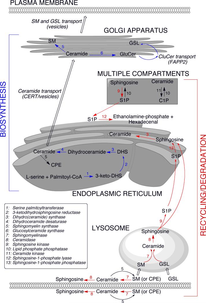
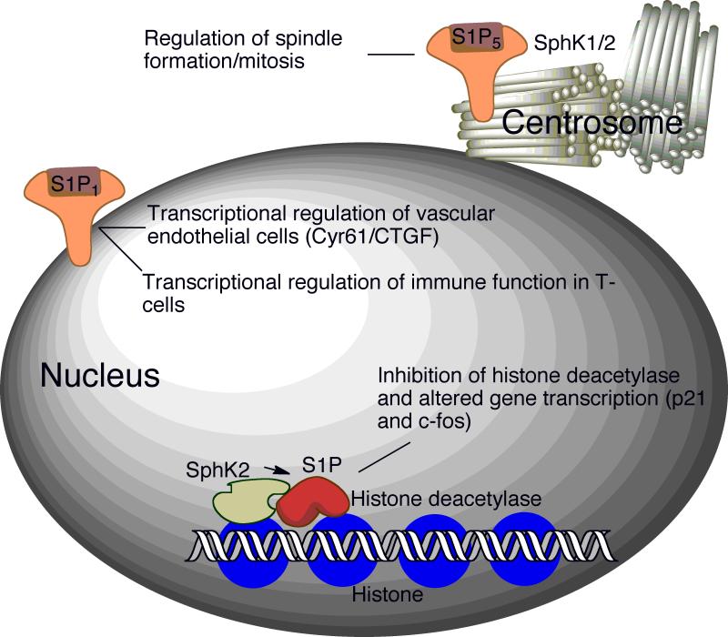
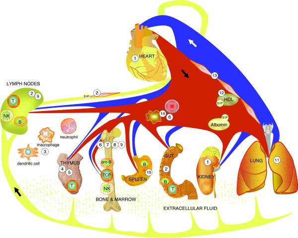
Sphingolipids comprise a complex family of naturally occurring molecules that are enriched in lipid rafts and contribute to their unique biochemical properties. Membrane sphingolipids also serve as a reservoir for bioactive metabolites including sphingosine, ceramide, sphingosine-1-phosphate and ceramide-1-phosphate. Among these, sphingosine-1-phosphate has emerged as a central regulator of mammalian biology. Sphingosine-1-phosphate is essential for mammalian brain and cardiac development and for maturation of the systemic circulatory system and lymphatics. In addition, sphingosine-1-phosphate contributes to trafficking and effector functions of lymphocytes and other hematopoietic cells and protects against various forms of tissue injury. However, sphingosine-1-phosphate is also an oncogenic lipid that promotes tumor growth and progression. Recent preclinical and clinical investigations using pharmacological agents that target sphingosine-1-phosphate, its receptors and the enzymes required for its biosynthesis and degradation demonstrate the promise and potential risks of modulating sphingosine-1-phosphate signaling in treatment strategies for autoimmunity, cancer, cardiovascular disease and other pathological conditions.


Figures

Figure 1
Figure 1. The sphingolipid metabolic pathway
The de novo biosynthesis of sphingolipids initiates in the endoplasmic reticulum (ER) where serine palmitoyltransferase condenses L-serine and palmitoyl-CoA, generating 3-ketodihydrosphingosine (3-keto-DHS), which is rapidly reduced to form dihydrosphingosine (DHS). DHS is N-acylated to form dihydroceramide, which is further converted into ceramide by the introduction of a double bond in the DHS base. Newly synthesized ceramide is transported to the Golgi apparatus where it is converted to sphingomyelin (SM) and glucosylceramide (GluCer). Ceramide transport to the Golgi is facilitated by CERT as well as vesicular transport . Transfer of GluCer from the cytoplasmic to the luminal side of the Golgi for the synthesis of complex glycosphingolipids (GSL) is facilitated by FAPP2 . SM and GSL are transported via vesicles from the Golgi to the plasma membrane. The recycling/degradation of higher order sphingolipids in the plasma membrane or lysosomal compartment also gives rise to ceramide, which can be further deacylated to yield sphingosine . Sphingosine and ceramide can be phosphorylated and de-phosphorylated by kinase and phosphatase activities, which exist in multiple compartments (see text for details). Sphingosine-1-phosphate (S1P) can be irreversibly degraded by S1P lyase located at the cytoplasmic side of the ER. Alternatively, S1P can be de-phosphorylated by specific phosphatases at the luminal side and converted back to ceramide for recycling (salvage pathway). The salvage pathway may also utilize sphingosine exiting directly from the lysosome. SM synthase activity resulting in ceramide-phosphoethanolamine (CPE) formation is found in the plasma membrane and ER.
Figure 2
Figure 2. Sphingodynamics
A sphingodynamics model represents the sum of balanced and unbalanced forces exerted upon cells via the interplay between bioactive sphingolipids and their targets in cells, membranes, tissues and circulating fluids. Cer = ceramide; So = sphingosine; SD = sphingadiene.
Figure 3
Figure 3. S1P signaling in the nucleus
Recently, nuclear functions of S1P have been revealed. Signaling through nuclear-associated S1P1 regulates gene transcription in endothelial cells and T-cells, thereby affecting vasculature and immune functions ,. S1P formed by SphK2 inhibits histone deacetylases. Complexes containing SphK2 and histone deacetylases are enriched in the promotor regions of p21 and c-fos genes . S1P5 and both SphK isoforms are found within the centrosome, where they have been postulated to play a role in spindle formation and mitosis .
Figure 4
Figure 4. S1P signaling in immunity and tissue injury
S1P signaling contributes to many aspects of innate and adaptive immunity. Pharmacological modulation of this pathway may attenuate tissue injury from autoimmune diseases, ischemia, sepsis and other conditions. 1) S1P/S1P1,2,3 mediates cardioprotection. Pharmacological activation of S1P1 and/or inhibition of SPL protect against cardiac and renal ischemia/reperfusion injury. 2) Lymph S1P is generated by lymphatic endothelium. This S1P source is required for lymphatic patterning and lymphocyte egress from lymph nodes and Peyer's patches. FTY720-phosphate blocks lymphocyte trafficking through functional antagonism and prolonged downregulation of S1P1 . 3) S1P/S1P2 inhibits macrophage migration. S1P signaling contributes to trafficking/effector functions of bone marrow (BM)-derived cells of innate immune system. 4) S1P regulates thymic endothelial P-selectin expression, settling of TCPs and mature T cell egress at the thymic corticomedullary junction, where pericyte-generated S1P guides S1P1-expressing thymocytes into the circulation. 5) SPL in the thymus helps establish S1P chemotactic gradients. 6) Complement activation induces release of S1P from erythrocytes, facilitating hematopoietic progenitor cell egress from BM. 7) S1P/S1P1 facilitates pre-B cell egress from BM into the circulation. 8) S1P/S1P1 regulates migratory behavior of osteoclast precursors (relevant to pathophysiology of rheumatoid arthritis and osteoporosis). 9) S1P/S1P1,5 facilitates NK cell egress from BM and lymph nodes. 10) S1P/S1P1 induces marginal zone B cell migration into follicles. 11) S1P/S1P1 and SPL inhibition reduce endotoxin-induced lung injury. 12) HDL-bound S1P may mediate protection from atherosclerosis. 13) S1P/S1P1,2,3 regulates vascular permeability and arterial pressure. 14) Erythrocytes and platelets generate blood S1P.

References

    1. Holthuis JC, Pomorski T, Raggers RJ, Sprong H, Van Meer G. The organizing potential of sphingolipids in intracellular membrane transport. Physiol Rev. 2001;81:1689–723. - PubMed
    1. Merrill AHJ, Sandhoff K. Sphingolipids: metabolism and cell signaling. In: Vance DE, Vance JE, editors. Biochemistry of Lipids, Lipoproteins and Membranes. Chapter 14; 2002. pp. 373–407.
    1. Mandala S, et al. Alteration of lymphocyte trafficking by sphingosine-1-phosphate receptor agonists. Science. 2002;296:346–9. - PubMed
    1. Brinkmann V, et al. The immune modulator FTY720 targets sphingosine 1-phosphate receptors. J Biol Chem. 2002;277:21453–7. - PubMed
    1. Lahiri S, Futerman A. The metabolism and function of sphingolipids and glycosphingolipids. Cell Mol Life Sci. 2007;64:2270–2284. - PMC - PubMed

Publication types

MeSH terms