Review
. 2010 Jul;11(7):515-21.
doi: 10.1038/embor.2010.92. Epub 2010 Jun 18.

Further assembly required: construction and dynamics of the endoplasmic reticulum network

Affiliations
Review

Further assembly required: construction and dynamics of the endoplasmic reticulum network

Seong H Park et al. EMBO Rep. 2010 Jul.

Abstract

The endoplasmic reticulum (ER) is a continuous membrane system comprising the nuclear envelope, ribosome-studded peripheral sheets and an interconnected network of smooth tubules extending throughout the cell. Although protein biosynthesis, transport and quality control in the ER have been studied extensively, mechanisms underlying the notably diverse architecture of the ER have only emerged recently; this review highlights these new findings and how they relate to ER functional specializations. Several protein families, including reticulons and DP1/REEPs/Yop1, harbour hydrophobic hairpin domains that shape high-curvature ER tubules and mediate intramembrane protein interactions. Members of the atlastin/RHD3/Sey1 family of dynamin-related GTPases mediate the formation of three-way junctions that characterize the tubular ER network, and additional classes of hydrophobic hairpin-containing ER proteins interact with and remodel the microtubule cytoskeleton. Flat ER sheets have a different complement of proteins implicated in shaping, cisternal stacking and microtubule interactions. Finally, several shaping proteins are mutated in hereditary spastic paraplegias, emphasizing the particular importance of proper ER morphology and distribution for highly polarized cells.


Conflict of interest statement

The authors declare that they have no conflict of interest.

Figures

Figure 1
Figure 1
Morphology of the endoplasmic reticulum. (A) A COS7 cell was immunostained with the ER markers Sec61β and DP1/REEP5 to reveal the different domains, as labelled. (B) Schematic diagram showing the domain organization of the ER, with tubular and sheet morphologies emphasized below. DP1, deleted in polyposis locus 1; ER, endoplasmic reticulum; REEP5, receptor expression-enhancing protein 5.
Figure 2
Figure 2
Schematic diagram of the tubular endoplasmic reticulum. Left: ER-shaping reticulon and DP1/REEPs/Yop1 proteins form large oligomers that shape ER tubules (Shibata et al, 2009). Atlastin/RHD3/Sey1 proteins are shown at three-way junctions. Right: Proposed membrane topologies for proteins involved in shaping the tubular ER network. DP1, deleted in polyposis locus 1; ER, endoplasmic reticulum; REEP, receptor expression-enhancing protein; RHD3, root hair defective 3; Rtn, reticulon; Sey1, synthetic enhancer of Yop1. Adapted with permission from Park et al (2010).
Figure 3
Figure 3
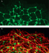
Interdependence of endoplasmic reticulum tubules and microtubules. (A) Atlastin 1 GTPase overexpressed in COS7 cells shows punctate enrichment along ER tubules (green) in the cell periphery, including at three-way junctions. (B) Microtubules are identified by co-immunostaining for β-tubulin (red). ER, endoplasmic reticulum. Adapted with permission from Park et al (2010).
Figure 4
Figure 4
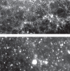
In vitro generation of a tubular endoplasmic reticulum network. (A) An ER network can be generated from a Xenopus microsomal fraction on addition of ATP and GTP in the absence of microtubules. (B) Network formation is inhibited by neutralizing atlastin antibodies. Similar inhibition is seen with neutralizing antibodies against reticulons and DP1/REEPs, but not against ER proteins such as IP3 receptor (Voeltz et al, 2006; Park et al, 2010). DP1, deleted in polyposis locus 1; ER, endoplasmic reticulum; IP3, inositol trisphosphate; REEP, receptor expression-enhancing protein. Images are courtesy of Dr Peng-Peng Zhu.

Similar articles

Cited by

References

    1. Anderson DJ, Hetzer MW (2008) Reshaping of the endoplasmic reticulum limits the rate for nuclear envelope formation. J Cell Biol 182: 911–924 - PMC - PubMed
    1. Andrade J, Zhao H, Titus B, Timm Pearce S, Barroso M (2004) The EF-hand Ca2+-bindng protein p22 plays a role in microtubule and endoplasmic reticulum organization and dynamics with distinct Ca2+-binding requirements. Mol Biol Cell 15: 481–496 - PMC - PubMed
    1. Barlowe C (2009) Atlastin GTPases shape up ER networks. Dev Cell 17: 157–158 - PubMed
    1. Baumann O, Walz B (2001) Endoplasmic reticulum of animal cells and its organization into structural and functional domains. Int Rev Cytol 205: 149–214 - PubMed
    1. Connell JW, Lindon C, Luzio JP, Reid E (2009) Spastin couples microtubule severing to membrane traffic in completion of cytokinesis and secretion. Traffic 10: 42–56 - PMC - PubMed

Publication types

MeSH terms