Review
. 2010 May 10;8(5):1650-80.
doi: 10.3390/md8051650.

On the chemistry, toxicology and genetics of the cyanobacterial toxins, microcystin, nodularin, saxitoxin and cylindrospermopsin

Affiliations
Review

On the chemistry, toxicology and genetics of the cyanobacterial toxins, microcystin, nodularin, saxitoxin and cylindrospermopsin

Leanne Pearson et al. Mar Drugs. .

Abstract

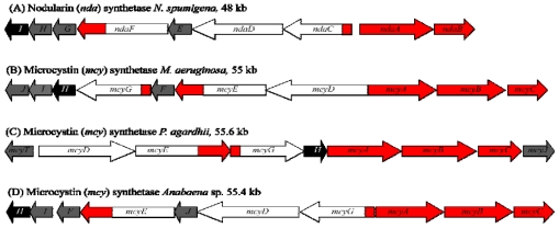
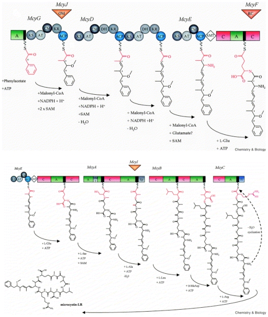
The cyanobacteria or "blue-green algae", as they are commonly termed, comprise a diverse group of oxygenic photosynthetic bacteria that inhabit a wide range of aquatic and terrestrial environments, and display incredible morphological diversity. Many aquatic, bloom-forming species of cyanobacteria are capable of producing biologically active secondary metabolites, which are highly toxic to humans and other animals. From a toxicological viewpoint, the cyanotoxins span four major classes: the neurotoxins, hepatotoxins, cytotoxins, and dermatoxins (irritant toxins). However, structurally they are quite diverse. Over the past decade, the biosynthesis pathways of the four major cyanotoxins: microcystin, nodularin, saxitoxin and cylindrospermopsin, have been genetically and biochemically elucidated. This review provides an overview of these biosynthesis pathways and additionally summarizes the chemistry and toxicology of these remarkable secondary metabolites.

Keywords: alkaloid; cyanotoxin; non-ribosomal peptide; polyketide; toxicology.


Figures

Figure 1
Figure 1
Structure of microcystin. General numbering of residues is indicated. Microcystin is a cyclic heptapeptide. The two variable amino acids in microcystin are indicated by X and Y. The most common isoform is microcystin-LR (MW 995.17), where X is l-Leu and Y is l-Arg.
Figure 2
Figure 2
Hepatotoxin gene clusters from various cyanobacteria. Structures of the microcystin and nodularin gene clusters of (A) N. spumigena, (B) M. aeruginosa, (C) P. agardhii, and (D) Anabaena sp. 90, showing genes encoding polyketide synthases (white), non-ribosomal peptide synthetases (red), tailoring enzymes (grey), and ABC-transporters (black). Diagram not drawn to scale.
Figure 3
Figure 3
(uper) Model for the formation of Adda during microcystin biosynthesis and predicted domain structure of McyG, McyD and McyE. (lower) Biosynthetic model for microcystin-LR and predicted domain structure of McyE, McyA, McyB, and McyC. Each circle and rectangle represents, respectively, a PKS or NRPS enzymatic domain. The aminotransferase domain is represented by a diamond. The activity of the tailoring ORFs, McyJ, F and I, are shown as inverted triangles. Abbreviations are as follows: A, aminoacyl adenylation; ACP, acyl carrier protein; AMT, aminotransferase; AT, acyltransferase; C, condensation; CM, C-methyltransferase; DH, dehydratase; Ep, epimerization; KR, ketoacyl reductase; KS, β-ketoacyl synthase; NM, N-methyltransferase; OM, O-methyltransferase; RC, racemase; TE, thioesterase. The NRPS thiolation motif is shown in black (reproduced from [22]).
Figure 4
Figure 4
Structure of nodularin. General numbering of residues is indicated. Nodularin is a cyclic pentapeptide (MW 619). The l-Arg residue of nodularin may be replaced with a homoarginine (nodularin-Har) or valine residue (motuporin).
Figure 5
Figure 5
(uper). Model of the formation of Adda during nodularin biosynthesis and predicted domain structure of NdaC, D and F. (lower). Biosynthetic model for nodularin and predicted domain structure of NdaF, H, A and B. Each grey and green circle represents, respectively, a PKS or NRPS enzymatic domain. The activities of the tailoring ORFs, NdaE, G and H, are shown as inverted triangles. Abbreviations are as follows: A, aminoacyl adenylation; ACP, acyl carrier protein; AMT, aminotransferase; AT, acyltransferase; C, condensation; CM, C-methyltransferase; DH, dehydratase; Ep, epimerization; KR, ketoacyl reductase; KS, β-ketoacyl synthase; NM, N-methyltransferase; OM, O-methyltransferase; RC, racemase; TE, thioesterase. (reproduced from [71]).
Figure 6
Figure 6
The core chemical structure of the paralytic shellfish poison (PSP), saxitoxin. ‘R’ represents variable positions. For a detailed list of isoforms see [98].
Figure 7
Figure 7
Structure of the paralytic shellfish toxin biosynthesis cluster (sxt) from; (a) Aphanizomenon sp. NH-5, (b) Anabaena circinalis AWQC131C, (c) Cylindrospermopsis raciborskii T3. Scale indicates gene length in kilobase pairs. Full bars and the letters A–E indicate common features between the various sxt gene clusters.
Figure 8
Figure 8
Proposed saxitoxin biosynthetic pathway in cyanobacteria based on intermediate characterization and bioinformatic analysis. Dashed lines indicate possible alternative reactions [see text for detailed steps].
Figure 9
Figure 9
The chemical structure of the NRPS and PKS derived alkaloid cylindrospermopsin. Natural variants containing an epimer at the hydroxyl bridge (C7) or lacking the hydroxyl altogether have also been reported [151].
Figure 10
Figure 10
Structural organization of the cylindrospermopsin gene cluster from C. raciborskii AWT205. Scale indicates gene cluster in base pairs.
Figure 11
Figure 11
Proposed biosynthetic pathway for cylindrospermopsin (see text for detail).

Similar articles

Cited by

References

    1. Sivonen K, Jones G. Toxic Cyanobacteria in Water: A Guide to Their Public Health consequences, Monitoring and Management. Vol. 1. E and FN Spon; New York, NY, USA: 1999. pp. 40–111.
    1. Welker M, von Dohren H. Cyanobacterial peptides -nature’s own combinatorial biosynthesis. FEMS Microbiol Rev. 2006;30:530–563. - PubMed
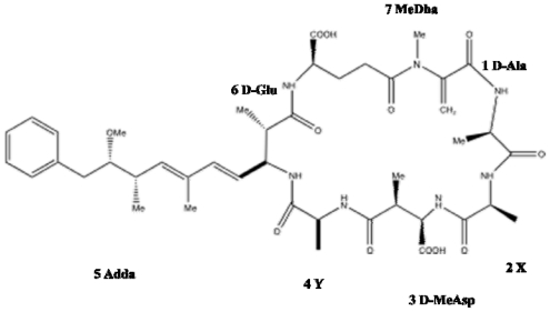
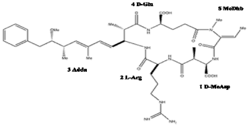
    1. Botes D, Wessels P, Kruger H, Runnegar M, Santikarn S, Smith R, Barna J, Williams D. Structural studies on cyanoginosins-LR, -YR, -YA, and -YM, peptide toxins from Microcystis aeruginosa. J Chem Soc. 1985;1:2747–2748.
    1. Namikoshi M, Yuan M, Sivonen K, Carmichael WW, Rinehart KL, Rouhiainen L, Sun F, Brittain S, Otsuki A. Seven new microcystins possessing two l-glutamic acid units, isolated from Anabaena sp. strain 186. Chem Res Toxicol. 1998;11:143–149. - PubMed
    1. Rinehart K, Namikoshi N, Choi B. Structure and biosynthesis of toxins from blue-green algae (cyanobacteria) J App Phycol. 1994;6:159–176.

MeSH terms

LinkOut - more resources