. 2010 Jun 15;5(6):e10919.
doi: 10.1371/journal.pone.0010919.

Supramolecular organization of the repetitive backbone unit of the Streptococcus pneumoniae pilus

Affiliations

Supramolecular organization of the repetitive backbone unit of the Streptococcus pneumoniae pilus

Glen Spraggon et al. PLoS One. .

Abstract

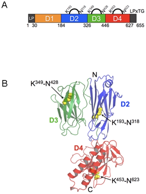
Streptococcus pneumoniae, like many other Gram-positive bacteria, assembles long filamentous pili on their surface through which they adhere to host cells. Pneumococcal pili are formed by a backbone, consisting of the repetition of the major component RrgB, and two accessory proteins (RrgA and RrgC). Here we reconstruct by transmission electron microscopy and single particle image reconstruction method the three dimensional arrangement of two neighbouring RrgB molecules, which represent the minimal repetitive structural domain of the native pilus. The crystal structure of the D2-D4 domains of RrgB was solved at 1.6 A resolution. Rigid-body fitting of the X-ray coordinates into the electron density map enabled us to define the arrangement of the backbone subunits into the S. pneumoniae native pilus. The quantitative fitting provide evidence that the pneumococcal pilus consists uniquely of RrgB monomers assembled in a head-to-tail organization. The presence of short intra-subunit linker regions connecting neighbouring domains provides the molecular basis for the intrinsic pilus flexibility.


Conflict of interest statement

Competing Interests: M.S., E.M., M.B., N.N., C.E., M.A.B., M.H., R.R., A.C., V.M. and I.F. are employees of Novartis Vaccines and Diagnostics s.r.l. which funded the work. R.R., A.C., M.A.B., and V.M. hold company stock options. All other authors report no potential conflicts. No other relevant declaration relating to employment, consultancy, patents, products in development or policies on sharing data and materials is needed. All authors confirm that this does not alter their adherence to all the PLoS ONE policies on sharing data and materials.

Figures

Figure 1
Figure 1. Negative stain EM of purified S. pneumoniae pili.
(A) The micrograph shows a network of elongated thin structures indicated by white arrows. (B) Set of raw images of purified S. pneumoniae pili. (C) Representative class averages of purified S. pneumoniae pili segments. (D) Re-projection of the 3D reconstruction of the pilus. Scale bar in panel A 20 nm, scale bar in panels B, C and D 10 nm.
Figure 2
Figure 2. Three-dimensional reconstruction of the S. pneumoniae pilus.
Surface representation of the S. pneumoniae pilus viewed along three orthogonal directions. The reconstruction is rendered at the molecular mass expected for two RrgB subunits (66 kD each). Structure is viewed along three orthogonal directions. Surface representation rendered with Chimera (Pettersen EF et al 2004).
Figure 3
Figure 3. Structure of RrgB.
A) Sequence organization of the full-length RrgB protein. D1 is depicted in yellow, while D2, D3 and D4 domains are respectively in blue, green and red. Position of the intra-molecular isopeptide bonds, the leader peptide (LP) and LPXTG motif are indicated. B) Ribbon Diagram showing the three domains of RrgB coloured as in A. Isopeptide bonds in each domain are shown as yellow spheres. Figure produced with Pymol (http://www.pymol.org/).
Figure 4
Figure 4. Isopeptide Bonds of RrgB and Spy0128.
A) and B) the isopeptide bond between residues Lys193 and Asn318 in domain 2 (D2) of RrgB. Dashed lines show the distance of the carboxylate anion provided by Asp241; the isopeptide bond between Lys 349 and Asn428 in domain 3 (D3) and the position of Glu405 contributing the carboxylate anion; the isopeptide bond between Lys 453 and Asn623 and the position of Glu577 in domain 4 (D4). C) Sigma-a weighted 2FoFc map contoured at 1σ level is shown for the isopeptide bond in D4 of RrgB. D) Isopeptide bond between Lys36 and Asn168 in Spy0128 (drawn from PDB 3b2m). Carbon atoms are coloured green for residues involved in the formation of the isopeptide bonds, yellow for neighbouring hydrophobic residues, and gray for residues adjacent to the isopeptide bond. Oxygen and nitrogen atoms are coloured red and blue, respectively. Figure produced by Pymol.
Figure 5
Figure 5. Sequence alignments of related pili proteins from Gram-positive bacteria.
A) Sequence alignment showing intra-molecular isopeptide bond formation which involves conserved residues Lysine (K), Aspartic Acid (D), Glutamic Acid (E) and Asparagine (N). Residues are highlighted and coloured according to the domain localization. UniProtKB/TrEMBL accession codes are shown; RrgB-TIGR4, RrgB-6BSP and RrgB-23FTW represent the sequences of the three RrgB alleles of S. pneumoniae; SAG0645 (also known as GBS80) belongs to PI-1 of Streptococcus agalactiae; GBS1477, SAL1486, SAG1407 and SAI1511 belong to GBS PI-2a, whereas SAK0776 belongs to GBS PI-2b; Spy0116 belongs to the M4 serotype of Streptococcus pyogenes. The sequence alignment was obtained and edited by Jalview/ClustalW . B) Cartoon representation of RrgB single domains coloured as in 1A) is shown on the bottom. Yellow spheres depict the residues forming the isopeptide bonds.
Figure 6
Figure 6. Fitting of the RrgBD2–D4 crystal structure and of the RrgBD1 computer model into the 3D map of the S. pneumoniae pilus.
A) Semi transparent rendered surface representations of the 3-D map of the S. pneumoniae pilus viewed along the z-axis tilted by 90° showing the docking of two copies of the atomic resolution RrgBD2–D4 subunit into the pilus reconstruction. Empty densities present above the two fitted RrgBD2–D4 copies have a volume of ∼21×e3 Å3 each. B) Same semi-transparent rendered surface representations as in panel A showing the docking into the pilus reconstruction of two copies of the RrgBD2–D4 subunit and contemporaneously of two computer model RrgBD1 domains. The docking reveals that the inter-subunit density (7 Å thick) present between the upper and lower subunits could accommodate the 8-residue C- terminal tail, not present in the RrgBD2–D4 crystal. Crystal structures are in cartoon representation and the three domains are coloured following the nomenclature of Figure 3. Figure produced with Pymol (http://www.pymol.org/). Surface representation and molecule rendered with Chimera .

Similar articles

Cited by

References

    1. Källström H, Islam M, Berggren PO, Jonsson AB. Cell signaling by the type IV pili of pathogenic Neisseria. Biol Chem. 1998;273:21777–21782. - PubMed
    1. Telford JL, Barocchi MA, Margarit I, Rappuoli R, Grandi G. Pili in Gram-positive pathogens. 2006;4:509–519. - PubMed
    1. Elena SF, Whittam TS, Winkworth CL, Riley MA, Lenski RE. Genomic divergence of Escherichia coli strains: evidence for horizontal transfer and variation in mutation rates. Int Microbiol. 2005;8:271–278. - PubMed
    1. Mattick JS. Type IV pili and twitching motility. Annu Rev Microbiol. 2002;56:289–314. - PubMed
    1. Wenyuan S, Hong S. Type IV Pilus-Dependent Motility and Its Possible Role in Bacterial Pathogenesis. Infection and Immunity. 2002;70:1–4. - PMC - PubMed

Publication types

Substances